Download
1 / 35
350 likes | 514 Views
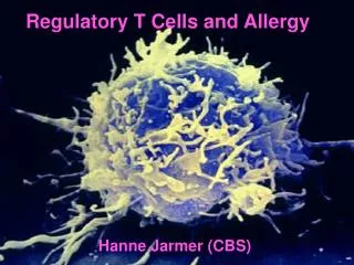
Regulatory T Cells and HIV Infection. ผศ. ศักดิ์ชัย เดชตรัยรัตน์ ดร. พานทอง สิงห์บุตรา ห้องปฏิบัติการเอชไอวี แขนงวิชาภูมิคุ้มกันวิทยาคลินิก คณะเทคนิคการแพทย์ มหาวิยาลัยเชียงใหม่. Immune Responses. Adaptive Immune Response. How this is achieved?. Self-Tolerance.

E N D
Regulatory T Cells and HIV Infection ผศ. ศักดิ์ชัย เดชตรัยรัตน์ ดร. พานทอง สิงห์บุตรา ห้องปฏิบัติการเอชไอวี แขนงวิชาภูมิคุ้มกันวิทยาคลินิก คณะเทคนิคการแพทย์ มหาวิยาลัยเชียงใหม่
Adaptive Immune Response How this is achieved?
Self-Tolerance • The immune system discriminates between self and non-self • Establishing and maintaining unresponsiveness to self • Central Tolerance (clonal deletion) • Occurs within central lymphoid organs • Before lymphocytes mature • Peripheral Tolerance • Occurs in peripheral lymphoid organs • After lymphocyte mature
Proposed Mechanisms of Peripheral Tolerance • Rendered anergic or further deleted if encounter self-antigen in the periphery • Fail to be activated because of low avidities to self-antigens • Lack of co-stimulation from APCs • Secluded from target self-antigens • Regulatory T cells actively down-regulate the activation and expansion of self-reactive lymphocytes
Regulatory T Cells (Tregs) • “Suppressor cells” (Gershonet al. 1972) • CD4+ T cells that express the IL-2 receptor α chain control autoreactive T cells in vivo (Sakaguchiet al. 1995) • Antigen-specific T-cell clones suppressed the proliferation of CD4+ T cells in response to antigen and prevented colitis in a severe combined immunodeficiency mouse model (Grouxet al., 1997)
Regulatory T Cells (Tregs) • Identification of several types of Tregs • “Regulatory T cells” is a preferred term
Types of Tregs Levi G., et al. Seminars in Immunology. 2011. in press.
CD4+CD25+Foxp3+ Regulatory T Cells • Fundamental in controlling various immune responses • Discovered by Sakaguchiet al. • 5-10% of peripheral CD4+ T cells • Expressed high level of IL-2Rα (CD25), Foxp3 • Developed in thymus and present in healthy individuals from birth
γδ Regulatory T Cells • Comprise 5% of total T cells in peripheral lymphoid tissues • Enriched in skin, intestine and genito-urinary tract
Natural Killer T Cells (NKT) • Produce large amount of Th1 (IFN-γ, TNF-α) and Th2 (IL-4, IL-10 and IL-13) cytokines • Play roles in tumor rejection, resistance to pathogenic infection, autoimmune diseases and allograft acceptance
Regulatory T Cells Type 1 (Tr1) • Arise in the periphery following activation of naïve T cells with Ag in the presence of IL-10 • Produce • high IL-10, TGF-β and IL-5 • low IL-2 and IFN-γ • no IL-4 • Do not express high levels of either CD25 or Foxp3
T Helper 3 (Th3) • Induced in gut environment with high TGF-β, Th2 cytokines, subsets of DC and oral antigens • Produces TGF-β • Induces tolerance to nonpathogenic resident bacteria and food antigens
CD8+ Regulatory T Cells • Arise either from thymus or in response to foreign or self antigens • CD8+CD25+ share similar phenotypes and functions with CD4+CD25+ T cells • Express increased mRNA levels of Foxp3, GITR, CCR8, TNFR-2 and CTLA-4 • Following activation, express TGF-β1 and CTLA-4, do not produce cytokines
Doble Negative T Cells (DN) • CD4-CD8-CD3+ comprised 1-2% of total CD3+ T cells in blood and lymph nodes • Express a unique set of cell surface markers • TCRαβ, CD25, LFA-1, CD69, CD45, CD30, CD62L and CTLA-4 • Produce • High IFN-γ • Low IL-10 and IL-4 • No IL-2
HIV Infection • Loss of CD4+ T cells • Chronic immune activation • Progressive immune disfunction • Impaired immune responses
Possible Roles of Treg in HIV Infection resistant to infection establish infection T cell T cell Quiescent Activated
Possible Roles of Treg in HIV Infection • destruction of T cells • deterioration of immune function T cell T cell Loss of Treg Hyperactivation • No protective immune responses • Establish carrier state of infection • Expand of Treg • Excessive Treg activity Suppressed
What are these markers? CD4 • A single chain molecule on T-helper cell • Binds to 2 domain of MHC-II • Increase the sinsitivity of T cell to Ag CD25 • Interleukin-2 receptor α-chain (IL-2Rα) • Receptor for IL-2, T-cell growth factor
What are these markers? Foxp3 • Forkhead ⁄ winged-helix transcription factor box P3 • Transcriptional repressor • Most specific Treg marker currently • Key factor in controlling Treg development
What are these markers? • Cytotoxic T lymphocyte associated protein-4 • Binds B7.1 (CD80)/B7.2 (CD86) • Major negative regulator of T-cell responses CTLA-4 (CD152)
What are these markers? GIRT • Glucocorticoid-induced tumor necrosis factor receptor • Required for the induction of apoptosis • Expressed on various lymphocytes at different levels • High surface expression of GITR is only confined to resting nTreg cells in the periphery and thymus
What are these markers? IL-7Rα (CD127) • Specific receptor chain for IL-7 • IL-7 control thymopoiesis and homeostasis of peripheral T lymphocytes
FACS profile of CD4+Foxp3+ Cells Foxp3 Foxp3 CD4 CD4 Isotypes control
Challenges in Identification of Tregs • Current markers are not truelyTreg-specific • All T cells express CD25 upon activation • CTLA-4 is upregulated on all CD4+ and CD8+ T cells, 2–3 days following activation • GITR is induced in T cells upon activation • CD127 is downregulated in most CD4+ T cells upon activation • Most human CD4+ and CD8+ T cells transiently express Foxp3 upon activation • Isolation of Treg using Foxp3 is limited to only phenotypic study