Download
1 / 89
890 likes | 1.14k Views
Assessment and Management of Patients with Endocrine Disorders. Dr Ibraheem Bashayreh. Location of the major endocrine glands. Definition of Hormones. Chemical messengers of the body Act on specific target cells Regulated by negative feedback Too much hormone, then hormone release reduced

E N D
Assessment and Management of Patients with Endocrine Disorders Dr Ibraheem Bashayreh
Definition of Hormones • Chemical messengers of the body • Act on specific target cells • Regulated by negative feedback • Too much hormone, then hormone release reduced • Too little hormone, then hormone release increased
Glands of the Endocrine System • Hypothalamus • Posterior Pituitary • Anterior Pituitary • Thyroid • Parathyroids • Adrenals • Pancreatic islets • Ovaries and testes
Hypothalamus • Sits between the cerebrum and brainstem • Houses the pituitary gland and hypothalamus • Regulates: • Temperature • Fluid volume • Growth • Pain and pleasure response • Hunger and thirst
Hypothalamus Hormones • Releasing and inhibiting hormones • Corticotropin-releasing hormone • Thyrotropin-releasing hormone • Growth hormone-releasing hormone • Gonadotropin-releasing hormone • Somatostatin-=-inhibits GH and TSH
Pituitary Gland • Sits beneath the hypothalamus • Termed the “master gland” • Divided into: • Anterior Pituitary Gland • Posterior Pituitary Gland
Anterior Pituitary Gland • Promotes growth • Stimulates the secretion of six hormones • Controls pigmentation of the skin
Anterior Pituitary Gland Hormones • Growth Hormone-- • Adrenocorticotropic hormone • Thyroid stimulating hormone • Follicle stimulating hormone—ovary in female, sperm in males • Luteinizing hormone—corpus luteum in females, secretion of testosterone in males • Prolactin—prepares female breasts for lactation
Posterior Pituitary Gland • Stimulates the secretion of two hormones • Promotes water retention
Posterior Pituitary Hormones • Antidiuretic Hormone • Oxytocin—contraction of uterus, milk ejection from breasts
Adrenal Cortex • Mineralocorticoid—aldosterone. Affects sodium absorption, loss of potassium by kidney • Glucocorticoids—cortisol. Affects metabolism, regulates blood sugar levels, affects growth, anti-inflammatory action, decreases effects of stress • Adrenal androgens—dehydroepiandrosterone and androstenedione. Converted to testosterone in the periphery.
Adrenal Medulla • Epinephrine and norepinephrine serve as neurotransmitters for sympathetic system
Thyroid Gland • Butterfly shaped • Sits on either side of the trachea • Has two lobes connected with an isthmus • Functions in the presence of iodine • Stimulates the secretion of three hormones • Involved with metabolic rate management and serum calcium levels
Thyroid • Follicular cells—excretion of triiodothyronine (T3) and thyroxine (T4)—Increase BMR, increase bone and calcium turnover, increase response to catecholamines, need for fetal G&D • Thyroid C cells—calcitonin. Lowers blood calcium and phosphate levels • BMR: Basal Metabolic Rate
Parathyroid Glands • Embedded within the posterior lobes of the thyroid gland • Secretion of one hormone • Maintenance of serum calcium levels
Parathyroid • Parathyroid hormone—regulates serum calcium
Pancreas • Located behind the stomach between the spleen and duodenum • Has two major functions • Digestive enzymes • Releases two hormones: insulin and glucagon
Pancreatic Hormones Insulin Glucagon—stimulates glycogenolysis and glyconeogenesis Somatostatin—decreases intestinal absorption of glucose
Insulin • Produced by the Beta cells in the islets of Langerhans • Regulates blood glucose levels • Mechanisms • Eases the active transport of glucose into muscle and fat cells • Facilitates fat formation • Inhibits the breakdown and movement of stored fat • Helps with protein synthesis
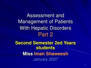
Action of insulin and glucagon on blood glucose levels. (A) High blood glucose is lowered by insulin release.
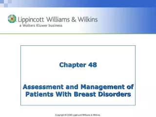
(continued) Action of insulin and glucagon on blood glucose levels. (B) Low blood glucose is raised by glucagon release.
Glucagon • Produced by the alpha cells in the islets of Langerhans • Glucagon released when blood glucose falls below 70 mg/dL
Glucagon • Prevents blood glucose from decreasing below a certain level • Functions: • Makes new glucose • Converts glycogen into glucose in the liver and muscles • Prevents excess glucose breakdown • Decreases glucose oxidation and increases blood glucose
Kidney • 1, 25 dihydroxyvitamin D—stimulates calcium absorption from the intestine • Renin—activates the RAS • Erythropoietin—Increases red blood cell production • RAS: Renin-Angiotensin System
Adrenal Glands • Pyramid-shaped organs that sit on top of the kidneys • Each has two parts: • Outer Cortex • Inner Medulla
Adrenal Cortex • Secretion of two hormones • Glucocorticoids: cortisol • Mineralocortocoids: aldosterone • Involved with blood glucose level, anti-inflammatory response, blood volume, and electrolyte maintenance
Adrenal Medulla • Secretion of two hormones • Epinephrine • Norepinephrine • Involved with the stress response
Ovaries • Estrogen • Progesterone—inportant in menstrual cycle,*maintains pregnancy,
Testes • Androgens, testosterone—secondary sexual characteristics, sperm production
Thymus • Releases thymosin and thymopoietin • Affects maturation of T lymphocetes
Pineal • Melatonin • Affects sleep, fertility and aging
Prostaglandins • Work locally • Released by plasma cells • Affect fertility, blood clotting, body temperature
Past Medical History • Hormone replacement therapy • Surgeries, chemotherapy, radiation • Family history: diabetes mellitus, diabetes insipidus, goiter, obesity, Addison’s disease, infertility • Sexual history: changes, characteristics, menstruation, menopause
Physical Assessment • General appearance • Vital signs, height, weight • Integumentary • Skin color, temperature, texture, moisture • Bruising, lesions, wound healing • Hair and nail texture, hair growth
Physical Assessment • Face • Shape, symmetry • Eyes, visual acuity • Neck
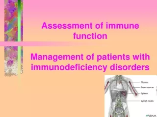
Palpating the thyroid gland from behind the client. (Source: Lester V. Bergman/Corbis)
Physical Assessment • Extremities • Hand and feet size • Trunk • Muscle strength, deep tendon reflexes • Sensation to hot and cold, vibration • Thorax • Lung and heart sounds • Extremity edema
Older Adults and Endocrine Function • Relationship unclear • Aging causes fibrosis of thyroid gland • Reduces metabolic rate • Contributes to weight gain • Cortisol level unchanged in aging
Abnormal Findings • Ask the client: • Energy level • Fatigue • Maintenance of ADL • Sensitivity to heat or cold • Weight level • Bowel habits • Level of appetite • Urination, thirst, salt craving
Abnormal Findings (continued) • Ask the client: • Cardiovascular status: blood pressure, heart rate, palpitations, SOB • Vision: changes, tearing, eye edema • Neurologic: numbness/tingling lips or extremities, nervousness, hand tremors, mood changes, memory changes, sleep patterns • Integumentary: hair changes, skin changes, nails, bruising, wound healing
Most Common Endocrine Disorders • Thyroid abnormalities • Diabetes mellitus
Diagnostic Tests • GH: fasting, well rested, not physically stressed • Water deprivation: fasting for 12 hours, no fluids/smoking after midnight • T3/T4: no specific preparation • Serum calcium/phosphate: fasting may or may not be required • Collection that needs to be iced or refrigerated
Diagnostic Tests • Cortisol/aldosterone level: two blood samples, client to be up for at least 2 hours before test is drawn • Urine 17-ketosteroids: 24-hour urine collection that needs to be iced or refrigerated
Diagnostic Tests • FBS: fast before the test • HbA1c: No fasting required • 2-hour OGTT: drink 75 g of glucose and do not eat anything until blood is drawn • Urine glucose/ketones: fresh urine specimen • Urine microalbumin: fresh urine specimen
Imaging Studies • MRI: metallic implants, lie motionless during test; remove all metal objects • CT scan: assess for allergies to iodine and seafood; lie immobile during the test • Thyroid scan: allergies to iodine and seafood; hold thyroid drugs containing iodine for weeks before the study
Imaging Studies • RAI: fast for 8 hours before; can eat 1 hour after radioiodine capsule/liquid taken; hold thyroid drugs with iodine for weeks before the study
THYROID DISORDERS Dr Ibraheem Bashayreh, RN, PhD
1-The gland as seen from the front is more nearly the shape of a butterfly. 2-composed of 2 encapsulated lobes, one on either side of the trachea, connected by a thin isthmus. 3-The thyroid extending from the level of the fifth cervical vertebra down to the first thoracic. The gland varies from an H to a U shape,overlying the second to fourth tracheal rings. 4-The pyramidal lobe is a narrow projection of thyroid tissue extending upward from the isthmus and lying on the surface of the thyroid cartilage. Thyroid Anatomy