Download
1 / 27
430 likes | 1.08k Views
Chapter 40 Assessment and Management of Patients With Biliary Disorders . Review of Anatomy and Physiology. Gallbladder Pancreas Insulin Glucagon Somatostatin. Liver, Biliary System, and Pancreas. Biliary Conditions:. Definition of terms: Biliary (chart 40-1)

E N D
Chapter 40Assessment and Management of Patients With Biliary Disorders
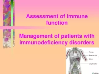
Review of Anatomy and Physiology • Gallbladder • Pancreas • Insulin • Glucagon • Somatostatin
Biliary Conditions: • Definition of terms: Biliary (chart 40-1) • Cholecystitis: Inflamation of the gallbladder • Cholelithiasis: the presence of calculi in the gallbladder • Cholecystectomy: removal of the gallbaldder • Cholecystostomy: opening and drainage of the gallbladder • Choledochotomy:opening into the common duct • Choledocholethiasis: stone in the common duct
Cont….. • Choledochlithotomy: incision of common bile duct for removal of stones • Choledochoduodenostomy: anastomosis of common duct to DU • Choledochojejunostomy: anastomosis of CD to jejunom • Lithotripsy: disintegration of gallstones by shock waves • Laparoscopic chlecystectomy: removal of gallbladder by endoscopic procedure • Laser Cholecystectomy: Removal of gallbladder using laser.
Cholelithiasis • Pathophysiology • Pigment stones precipitating of unconjugated pigment in the bile to form stones represent one third of cases. Causes cirrhosis, hemolysis and infection of the biliary tree • Cholesterol stones Cholesterol is insoluble in water, solubility depend on bile acid and lecithin (phospholipid) in bile. Decreased bile acid and increase cholesterol synthesis in liver cause bile supersaturation with cholesterol which precipitate and form stone Four X women than men • Risk factors multi para, Frequent changes in Wt, Rapid Wt loss, oral contraceptive, estrogens, cystic fibrosis, DM, and increases with age due to more cholesterol synthesis and decreased bile acid synthesis
Cholelithiasis—Manifestations • May have no or minimal symptoms and may be acute or chronic. • Epigastric distress: fullness, abdominal distention, vague upper right quadrant pain. Distress may occur after eating a fatty meal. • Acute symptoms occur with obstruction and inflammation or infection: fever, palpable abdominal mass, severe right abdominal that radiates to the back or right shoulder, nausea and vomiting. • Biliary colic is episodes of severe pain usually associated with nausea and vomiting, which usually occur several hours after a heavy meal. • Jaundice may develop due to blockage of the common bile duct.
Diagnostic Tests: • Abdominal X-ray • U/S • Radionuclide imaging: IV radioactive agent • Cholecystography: oral iodine contrast agent used 10-12 hrs before X-ray, NPO • Endoscopic retrograde cholengiopancretography (ERCP): Direct observation through fiberoptic scope inserted through esophagus into DU ( Discuss nursing implication). • Percutaneous transhepatic Cholengiography: inject the dye directly into the biliary tree.
Medical Management of Cholelithiasis • Cholecystectomy • Laparoscopic cholecystectomy • Dietary management • Medications: ursodeoxycholic acid and chenodeoxycholic acid • Nonsurgical removal • By instrumentation • Intracorporeal or extracorporeal lithotripsy
Nursing Process: The Care of the Patient Undergoing Surgery for Gallbladder Disease—Assessment • Patient history • Knowledge and teaching needs • Respiratory status and risk factors for respiratory complications postoperative • Nutritional status • Monitor for potential bleeding • Gastrointestinal symptoms: after laparoscopic surgery asses for loss of appetite, vomiting, pain, distention, fever—potential infection or disruption of GI tract
Nursing Process: The Care of the Patient Undergoing Surgery for Gallbladder Disease—Diagnoses • Acute pain • Impaired gas exchange • Impaired skin integrity • Imbalanced nutrition • Deficient knowledge
Collaborative Problems/Potential Complications • Bleeding • Gastrointestinal symptoms • Complications as related to surgery in general: atelectasis, thrombophlebitis
Nursing Process: The Care of the Patient Undergoing Surgery for Gallbladder Disease—Planning • Goals may include relief of pain, adequate ventilation, intact skin, improved biliary drainage, optimal nutritional intake, absence of complications, and understanding of self-care routines.
Postoperative Care Interventions • Low Fowler’s position • May have NG • NPO until bowel sounds return, then a soft, low-fat, high-carbohydrate diet postoperatively • Care of biliary drainage system • Administer analgesics as ordered and medicate to promote/permit ambulation and activities, including deep breathing • Turn, and encourage coughing and deep breathing, splinting to reduce pain • Ambulation
Patient Teaching—See Chart 40-3 • Medications • Diet: at discharge, maintain a nutritious diet and avoid excess fat. Fat restriction is usually lifted in 4–6 weeks. • Instruct in wound care, dressing changes, care of T-tube • Activity • Instruct patient and family to report signs of gastrointestinal complications, changes in color of stool or urine, fever, unrelieved or increased pain, nausea, vomiting, and redness/edema/signs of infection at incision site
Pancreatitis • A severe disorder that can lead to death. Acute pancreatitis does not usually lead to chronic pancreatitis. • Acute pancreatitis: the pancreatic duct becomes obstructed and enzymes back up into the pancreatic duct, causing auto digestion and inflammation of the pancreas. • Chronic pancreatitis: a progressive inflammatory disorder with destruction of the pancreas. Cells are replaced by fibrous tissue, and pressure within the pancreas increases. Mechanical obstruction of the pancreatic and common bile ducts and destruction of the secreting cells of the pancreas occur.
Causes: • Biliary tract disease: Stones • Long term use of alcohol • Bacterial or viral infection • Blunt abd trauma, peptic ulcer, ischemic vascular disease • Hyperlipidemia, hypercalcemia • Use of corticosteriods, thiazide diuretics, and oralcontraceptivr • ERCP or surgeries near to pancreas.
Assessment and diagnostic findings • History of Abd pain • Serum amylase and Lipase • Increase WBC’s • X-Ray studies • Ultrasound • Abd CT-Scan • ERCP rarely used because patient is acutely ill.
Severe abdominal pain Patient appears acutely ill Abdominal guarding Nausea and vomiting Fever, jaundice, confusion, and agitation may occur Ecchymosis in the flank or umbilical area may occur May develop respiratory distress, hypoxia, renal failure, hypovolemia, and shock Recurrent attacks of severe upper abdominal and back pain accompanied by vomiting Weight loss Steatorrhea Manifestations Acute Chronic
Nursing Process: The Care of the Patient With Acute Pancreatitis—Assessment • Focus on abdominal pain and discomfort • Fluid and electrolyte status • Medications • Alcohol use • GI assessment and nutritional status • Respiratory status • Emotional and psychological status of patient and family; anxiety and coping
Nursing Process: The Care of the Patient With Acute Pancreatitis—Diagnoses • Acute pain • Ineffective breathing pattern • Imbalanced nutrition • Impaired skin integrity
Collaborative Problems/Potential Complications • Fluid and electrolyte disturbances • Necrosis of the pancreas • Shock • Multiple organ dysfunction syndrome • DIC
Nursing Process: The Care of the Patient With Acute Pancreatitis—Planning • Major goals include relief of pain and discomfort, improved respiratory function, improved nutritional status, maintenance of skin integrity, and absence of complications.
Relieving Pain and Discomfort • Use of analgesics • Nasogastric suction to relieve nausea and distention • Frequent oral care • Bed rest • Measures to promote comfort and relieve anxiety