Download
1 / 1
10 likes | 111 Views
**. 3. 2. PP1 Activity (x10 6 U/g protein). **. **. **. 1. 0. C 6 - Cer. control. 5. 10. 20. 30. 40. C 8 -GlcCer ( M). A Novel Lipid Inhibitor of Protein Phosphatase-1

E N D
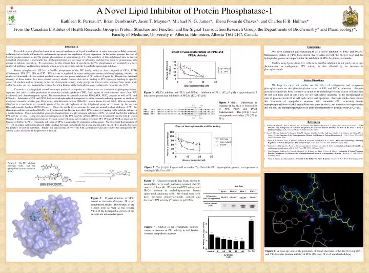
** 3 2 PP1 Activity (x106 U/g protein) ** ** ** 1 0 C6- Cer control 5 10 20 30 40 C8-GlcCer (M) A Novel Lipid Inhibitor of Protein Phosphatase-1 Kathleen R. Perreault*, Brian Dembinski^, Jason T. Maynes*, Michael N. G. James*, Elena Posse de Chaves^, and Charles F. B. Holmes* From the Canadian Institutes of Health Research, Group in Protein Structure and Function and the Signal Transduction Research Group, the Departments of Biochemistry* and Pharmacology^, Faculty of Medicine, University of Alberta, Edmonton, Alberta T6G 2H7, Canada Introduction Reversible protein phosphorylation is an integral mechanism of signal transduction in many important cellular processes, including, but certainly not limited to, mitogenesis, apoptosis, and regulation of gene expression. In the human genome, the ratio of Ser/Thr protein kinases to Ser/Thr protein phosphatases is approximately 8:1. The corollary of this unbalanced ratio is that an individual phosphatase is responsible for dephosphorylating a broad range of substrates, and therefore must be promiscuous with respect to substrate specificity. To compensate for this relative lack of specificity, Ser/Thr phosphatases are regulated by a large number of inhibitory and targeting subunits, which serve to direct their activity towards the appropriate substrate. Protein phosphatase-1 (PP1) is a Ser/Thr phosphatase of the PPP family, which is also comprised of PP2A, PP2B (Calcineurin), PP4, PP5, PP6 and PP7. PP1 activity is regulated by many endogenous protein inhibiting/targeting subunits. A number of structurally diverse natural product toxins are also potent inhibitors of PP1 activity (Figure 1). Despite the structural diversity of these toxins, they have several coarsely similar features that aid in binding to PP1: hydrogen bonding to specific conserved residues in close proximity to the site of enzymatic activity, acidic groups that interact with conserved basic amino acids within the active site, and hydrophobic regions that allow binding at the hydrophobic groove adjacent to the active site (1-3). Ceramide is a sphingolipid second messenger produced in response to cellular stress via activation of sphingomyelinases. Agonists that cause cellular production of ceramide include cytokines (TNF, Fas), agents of environmental stress (heat, UV irradiation), and chemotherapeutic agents. The accumulation of ceramide activates JNK/SAPK, PKCζ, caspases as well as PP1 and PP2A (6). Substrates of PP1 and PP2A that are dephosphorylated in response to either ceramide-inducing agonists or addition of exogenous ceramide include c-jun, SR proteins, retinoblastoma protein, PKB/Akt1, protein kinase Cα and Bcl-2. Glucosylceramide (GlcCer) is a metabolite of ceramide produced by the glycosylation of the 1-hydroxyl group of ceramide by the enzyme Glucosylceramide Synthase (GCS) (Figure 1). Given the similarities in structure between the natural product inhibitors of PP1, the clavosines, and the sphingolipid GlcCer, we hypothesized that GlcCer may affect PP1c activity by binding to the catalytic subunit in a similar fashion. Using radiolabelled glycogen phosphorylase a, a physiological substrate of PP1, we found that GlcCer inhibited PP1 activity in vitro. Using site-directed mutagenesis of the PP1 catalytic subunit (PP1c), we determined that the β12-β13 loop (Figures 2 and 4), an unstructured chain of five non-conserved amino acid residues present in PP1, PP2A and PP2B, is important for binding of GlcCer to PP1c. Ceramide activation of PP1c is unaffected by mutations in this region. We also found that mutation of Tyr-134, an amino acid residue present at the interface between the hydrophobic groove and the active site of PP1c, greatly decreases the potency of GlcCer inhibition. Finally, we used lysates of live cells with accumulated GlcCer to show that endogenous PP1 activity is also decreased in the presence of GlcCer. Conclusions We have identified glucosylceramide as a novel inhibitor of PP1c and PP2Ac. Mutagenesis studies of PP1c have shown that residues in both the β12-β13 loop and the hydrophobic groove are important for the inhibition of PP1c by glucosylceramide. Studies using lysates from live cells show that this inhibition is not purely an in vitro phenomenon, as endogenous PP1 activity is also affected by an increase in glucosylceramide. Results Future Directions We hope to carry out studies on the effect of endogenous and exogenous glucosylceramide on the phosphorylation states of PP1 and PP2A substrates. Because glucosylceramide has been shown to accumulate in multidrug-resistant cancer cell lines like the KB cell lines used in our study, we are particularly interested in the phosphorylation state of proteins involved in cell cycle arrest and apoptosis. Previous studies have shown that treatment of sympathetic neurons with ceramide (PP1 activator) blocks hyperphosphorylation of pRB (retinoblastoma gene product), and therefore we hypothesize that we may see hyperphosphorylation of pRB upon treatment of neurons with GlcCer (5). Figure 3: GlcCer inhibits both PP1c and PP2Ac. Inhibition of PP1c (IC50~5 μM) is approximately 3 times more potent than inhibition of PP2Ac (IC50~15 μM). Figure 4 (left): Differences in sequence in the β12-β13 loop region of PP1, PP2A and PP2B (Calcineurin). The β12-β13 loop corresponds to residues 273-277 in PP1c. • References • 1. Kathleen R. Perreault, Jason T. Maynes, Maia M. Cherney, Hue Anh Luu, Michael N. G. James, and Charles F. B. Holmes. Crystal Structure and Mutagenesis of a Protein Phosphatase-1:Calcineurin Hybrid Elucidate the Role of the ß12-ß13 Loop in Inhibitor Binding.J. Biol. Chem. 279: 43198-43206 (October 2004). • 2. Jason T. Maynes, Katherine S. Bateman, Maia M. Cherney, Amit K. Das, Hue Anh Luu, Charles F. B. Holmes, and Michael N. G. James. Crystal Structure of the Tumor-promoter Okadaic Acid Bound to Protein Phosphatase-1.J. Biol. Chem. 276: 44078-44082 (November 2001). • 3. Charles F. B. Holmes, Jason T. Maynes, Kathleen R. Perreault, and Michael N. G. James. Molecular Enzymology Underlying Regulation of Protein Phosphatase-1 by Natural Toxins.Curr. Med. Chem. 9: 1981-1989 (November 2002). • 4. Yaakov Lavie, Hui-ting Cao, Stuart L. Bursten, Armando E. Giuliano, and Myles C. Cabot. Accumulation of glucosylceramides in multidrug-resistant cancer cells.J. Biol. Chem. 271:19530-6 (August 1996). • Greg Plummer, Kathleen R. Perreault, Charles F. B. Holmes, and Elena I. Posse de Chaves. Activation of Serine/Threonine Protein Phosphatase-1 is Required for Ceramide-Induced Survival of Sympathetic Neurons.Biochem. J.385: 685-693 (February 2005). • Yusuf A. Hannun and Chiara Luberto. Ceramide in the Eukaryotic Stress Response.Trends Cell Biol. 10: 73-80 (February 2000). Figure 1: The PP1 inhibitor clavosine (left), and the sphingolipid glucosylceramide (right). Figure 5: The β12-β13 loop as well as residue Tyr-134 of the PP1c hydrophobic groove, are important in binding of GlcCer to PP1c. Figure 6: Glucosylceramide has been shown to accumulate in several multidrug-resistant (MDR) cancer cell lines (4). We examined PP1 activity and GlcCer content in multidrug-resistant human epidermoid carcinoma cells. We found these cells have increased glucosylceramide content and decreased PP1 activity. (** refers to p<0.005). Cell Type KB-V.01 KB-V.1 KB-V1 KB-3-1 PP1 Activity (x107U/g protein) 4.6±0.3 4.0±0.2* 3.4±0.1** Figure 2: Crystal structure of PP1c bound to clavosine (Maynes, JT et al, unpublished results. The residues of the β12-β13 loop as well as the residue Y134 of the hydrophobic groove of the enzyme are indicated in green. 3.6±0.1** GluCer content (TLC) Figure 7: GlcCer in rat sympathetic neurons causes a decrease in PP1 activity in cell lysates from rat sympathetic neurons. Figure 8: A close-up view of the proximity of bound clavosine to the β12-β13 loop (left) and Y134 residue (bottom middle) of PP1c (Maynes, JT et al, unpublished data).