Download
1 / 11
110 likes | 247 Views
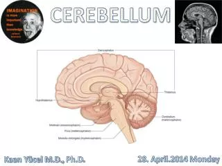
Cerebellum I. Introduction - Basic circuit is a loop between cerebral cortex, basal ganglia/cerebellum, and thalamus, with modulating input at second item - Anterior, posterior, flocculonodular lobes, primary and posterolateral fissures - Vermis , paravermis , lateral hemispheres. .

E N D
Introduction - Basic circuit is a loop between cerebral cortex, basal ganglia/cerebellum, and thalamus, with modulating input at second item - Anterior, posterior, flocculonodular lobes, primary and posterolateral fissures - Vermis, paravermis, lateral hemispheres.
I. VESTIBULOCEREBELLUM - Vestibulocerebellum = flocculonodular lobe - Input: vestibular nuclei and Scarpa’s (vestibular) ganglion - Output: vestibular nuclei to control head/eye movements (e.g VOR) - Clinical signs: impaired vestibular function, poor eye control, nystagmus.
II. SPINOCEREBELLUM - Spinocerebellum = vermis + paravermis - Vermisfastigial nucleus, paravermis interposed nuclei - Input - Clarke’s nucleus (via dorsal spinocerebellar tract) and lateral cuneate nucleus (via cuneocerebellar tract) provide ipsilateral muscle spindle
- Ventral spinocerebellar tract (deep tendon receptors). - Reticular formations via spinal input - Output - Vermisfastigial nucleus pontine reticular formation for extensor a
- Paravermis interposed nuclei red nucleus, VL thalamus for motor function - Clinical signs - Cerebellarhypotonia: reduced muscle tone due to reduced deep nuclei activity - Dysmetria: failure to make seamless coordination, muscle overshoots.
III. PONTOCEREBELLUM - Pontocerebellum = lateral hemispheres - Input: cerebral cortex via contralateralpontine nuclei - Output: dentate nucleus VL thalamus for motor planning and medial thalamus for working memory - Clinical signs: decomposition of movement into a series of individual acts separated by stops.
IV. Cerebellar circuitry - Mossy fibers: various origins granule cells; excites many PCs via parallel fibers - Climbing fibers: inferior olive Purkinje cells; powerful excitation - Purkinje cells: planar in stacks, input from many MFs and single CF, output to deep nuclei.
- Basket and stellate cells inhibit Purkinje cells whereas golgi cells inhibit granule cells - Spacing of PCs allows for precisely timed actions in response to rich, complex input - Circuitry is responsible for learning by practice.
V. Cerebellar Peduncles A) Inferior peduncle - Input from dorsal spinocerebellar tract, cuneocerebellar tract, olivocerebellar afferents - Some efferents to vestibular nuclei, but mostly input.
B) Middle peduncle - Input from pons to pontocerebellum C) Superior peduncle - Output to red nuclei and VL thalamus (latter is input to primary motor cortex) - Input from ventral spinocerebellar tract (minor).