Download
1 / 49
490 likes | 513 Views
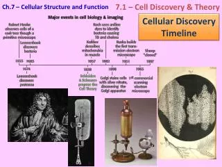
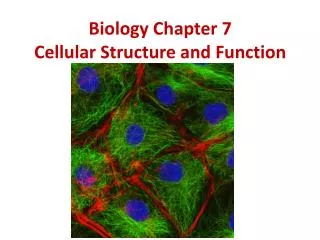
Cellular Structures and Function. History of the cell. Robert Hooke – English – first discovered cells, 1665-cork Anton von Leeuvenhoek – Dutch – pond water – animalcules (little animals) Matthias Schleiden – German botonist– cells plants, composed of units called cells

E N D
History of the cell Robert Hooke – English – first discovered cells, 1665-cork Anton von Leeuvenhoek – Dutch – pond water – animalcules (little animals) Matthias Schleiden – German botonist– cells plants, composed of units called cells Theodor Schwann - German zoologist – all animals compare
Modern Cell Theory • Cells are the smallest complete living things – basic units of organization • All organisms are composed of one or more cells in which all life process occur • Cells arise only from preexisting cells through the process of cell division • All of today’s existing cells are descendants of the first cells
Cells in general • 50-100 trillion cells • 260 cells varieties • Differentiated – cells with specialized characteristics, distinctive shape for function • Measured in mircrometer, 10-6 m
Composite cells • 3 parts of a cell: • Cell membrane – outermost limit, defines cell, thin, selectively permeable, signal transduction, message response • Cytoplasm • Nucleus (eurkaryotic) – typical, membrane bound, Prokayrotic – lacks, bacteria
Cell membrane, plasma membrane, plasmalemma Double phospholipid layer with proteins. Phosphorus – blue balloon, hydrophillic (attracts water) Lipid – purple tails, hydrophobic (repels water) Proteins form channels to allow passage of molecules (facilitated diffusion), pores Fluid mosaic pattern
Proteins • Integral – spans the cell membrane • Peripheral – projects from surface • Transmembrane – in cytoplasm project through cell membrane, coiled – receptors • Cellular adhesion – molecule (CAM) – cells touch / bind, glycoprotein – “self”
CAMS • Some cells must move, ex. White blood cells • Selectin – covers WBC, provide traction as nearing injury site • Integrin – allows WBC to stick to injury site where it will destroy bacteria.
Cytoplasm Protoplasm – liquid part (of cell) Cytoplasm (cytosoll)– outside nucleus Nucleoplasm – inside nucleus Water, solution – individual atom distributed throughout Colloid – clumps of atoms distributed though out medium Cytoskeleton – protein rod / tubules for support
Cellular Organelles • There are several reasons why cells evolved organelles. First, organelles can perform specialized functions (cilia, these short filaments act as "paddles" to help some cells move). • Second, membrane bound organelles can act as containers, separating parts of the cell from other parts of the cell. • Third, the membranes of organelles can act as sites for chemical reactions.
Cellular Organelles • Nonmembrane Bound Organelles- • Ribosome's • Centrioles • Microtubules • Membrane Bound Organelles- • Nucleus • Mitochondria • Lysosomes • Endoplasmic reticulum • Golgi Apparatus • Peroxisomes
Nucleus • Control center of the cell,double nuclear membrane (envelope). Nucleoplasm fluid in the nucleus. Membrane has pores to allow material out (mRNA). • Nucleic acid called chromatin found. They shorten and thicken during cell division. • Nucleolus – spherical particle in nucleoplasm where ribosomes are made (synthesized).
Mitochondria • Powerhouse of the cell. Composed of two membranes. Inner folds called cristae. Where anerobic cellular respiration occurs. Cells with higher energy requirements have more mitochondria (muscles) • DNA passed on by mother
Lysosomes • Small structures in cytoplasm surrounded by membrane and contain digestive enzymes. • Functions: digest stored foods (with vacoule), repair of organelles, suicide agents in old, weakened cells
Peroxisomes • Resembles lysosome • Found primarily in liver and kidney • Catalyzes metabolic reactions that release hydrogen peroxide (synthesize bile acids, breakdown lipid, degradation of rare biochemicals, alcohol detox
Endoplasmic reticulum • Membrane – bound channels called cisternae • Connects outer nuclear membrane and cell membrane, transports • Rough E.R. – attached ribosomes, protein synthesis • Smooth (agranular) E.R. - transports fats, synthesis of sex hormones.
Golgi apparatus, body, complex • Collection of flat saclike cisternae that look like stack of pancakes • Compounds to be secreted are concentrated and collected (wrapped), act as a warehouse • Carbohydrate synthesized if cell’s job
Ribosomes • Small granules in cytoplasm and attached to ER. (no membrane) • Proteins and RNA • Site of protein synthesis • Proteins are part of structure of membranes, enzymes or catalyst for reactions, immune response
Centrioles • Two centrioles are found at right angles to each other near nuclear membrane (pair as centrosome) • Each centriole is composed of 9 sets of triplet fibers • Centrioles form spindle fibers during cell division and guide the chromosomes to their daughter cells
Cillia and Flagella • Composed of fibrils • Cillia are short, many rows; move materials across the free surface of cell. (respiratory tract cells) • Flagellum are long, hair like, one or two, will propel the cell through a medium (sperm cell)
Microfilaments and microtubules • Distinguished by protein type, diameter and how they assemble • Microfilaments – tiny rods of protein actin, form meshwork provide cellular movement (myofibrils in muscle) • Microtubules – globular protein tubulin, rigid – cell shape, • Specialized cells have intermediate filaments which form dimers (protein pairs). Epidermis cells attach
Inclusions • Temporary structures • Stored nutrients – glycogen and lipids • Pigments - like melanin in skin
Plant cells vs. animal cells • Plant cells have a cell wall made of cellulose (fiber in food, not digested) • Chloroplast – green structure that are responsible for photosynthesis. • 6CO2 + 12 H2O -> C6H12O6 + 6O2 + 6 H2O • Chromoplasts – carotenoid pigments (Xanthophyll – yellow, carotene – orange) • Leucoplast – no pigment, storage plastids (onion bulb) • Vacuole – water storage
Process of Protein Synthesis • Code to make a protein is a gene on a DNA molecule. • Messenger RNA (mRNA) copies the code form DNA in a process called transcription. mRNA leaves the nucleus through a pore and takes the code to a ribosome or group of ribosomes • Transfer RNA (tRNA) go into the cytoplasm and pick up a particular amino acid. Each tRNA is coded for Amino acid by its anti-codon loop and will only match a particular site on the mRNA molecule called a codon. This process is called translation • Ribosome (rRNA) will now link up Amino acid brought to mRNA by the tRNA to form a protein.
Discovers of DNA (heredity material of cell) • Friedrich Miescher – 1869 – German chemist isolated the nucleic acid • P.A. Levine – 1920 – Discovered DNA contained Phosphates, five – carbon sugars and nitrogen bases. These form a nucleotide • Rosalind Franklin – British – discovered the helical structure using crystallography • James Watson – American – Francis Crick – British – won the 1962 Nobel Prize for working out the 3 dimensional structure (double helix).
Anatomy of a DNA molecule • Double helical chain (twisted ladder) of nucleotides • Nucleotides has phoshate group (PO4), Five – carbon sugar (deoxyribose), and a nitrogen – containing base (purine or pyrimidine) • Pyrimidine – single ring of 6 carbon atoms of C and N.(thymine or cytosine) • Purine – fused double ring of 9 atoms of C and N. (adenine and guanine) • Sugar and phosphate make the backbone. The bases attach to the sugar
The four nitrogen bases are, Adenine(A) Guanine(G) Thymine(T) Cytosin(C) Adenine bonds with Thymine Cytosin bonds with Guanine These double nitrogen-bases bind to form what is known as the “DNA Double Helix” The Four Nitrogen Based pairs
Mitosis • Cell cycleG1 – growth, S – synthesis, G2 growth, Mitosis- cellular division • Single cell duplicates itself –allows body to growth, repair, maintain structure; maintains our life • Mitosis is nuclear division plus cytokinesis- division of the cytoplasm and produces two identical daughter cells.
Prophase-chromatin shorten and darken to form chromosomes, nuclear membrane disappears, centrioles and spindle fibers form Metaphase- chromosomes line up at middle, spindle fibers attach to centromere and centrioles Anaphase- chromosome split and move towards poles (centrioles) Telophase- Chromosomes at the poles Cytokinesis – splitting of the cytoplasm (with organelles) Interphase is often included in discussions of mitosis, but interphase is technically not part of mitosis, but rather encompasses stages G1, S, and G2 of the cell cycle.
Meiosis • http://cellscienceproject.tripod.com/sitebuildercontent/sitebuilderfiles/meiosis.mov • http://www.trentu.ca/biology/101/14.html • Increases genetic variability – offspring a chance to adapt to changing environment • Occurs in ovaries – females, (oogenesis – ovum sex cell), testes – male (spermatogensis – sperm sex cell) • Reduction division of nuclear material – ½ (23 chromosomes) genetic material • Mutation – exact copy of genetic code is disrupted, producing a variation
Meiosis • Prophase 1 – Homologous chromosomes pair and crossing over may occur • Metaphase 1 – Homologous pairs align along equator • Anaphase 1 – Centromere do NOT divide. Each chromosome goes to pole • Telophase 1 – Division • Prophase 2 – Chromosome appears • Metaphase 2 – Chromosomes line up at equator • Anaphase 2 – Centromeres divide • Telephase 2 – Chromatids at pole, cytokinesis, 4 haploid cells
Control of cell division • Telemere – tip of chromosomes, same 6 nucleotides repeat hundreds of times, when tip wear down – cells stops dividing • Surface area to cell volume • Hormones and growth factors • Space availability – contact inhibition
Stem and progenitor cells • Differentiation – process of specialization • Stem cells – able to divide to give rise to other progenitor stem cells – “self renewing”; egg and embryo cells are totipotent – give rise to every cell type • Progenitor cells – partially specialized stem cells, continues specialization; pluripotent – daughter cells follow several pathways, but not all.
Cell death • Apoptosis – programmed cell death – normal part of development, remove excess skin (webbed digits) and peeling sunburn, orderly • Not the same as necrosis – cell death due to injury or inflammation • Cell membrane receives a signal to die – enzyme caspases cuts up components and cell membrane covers (blebs) and phagocytizes
Mutations • Change in DNA that effects a person, detectable • Most Single nucleotide polymorphisms – change in 1 base pair, no effects. • Spontaneous – chemical tendency of bases to exist in 2 slightly different ways. • Induced – exposed to a mutagen – agent that causes a mutation (radiation, chemicals. • If change is in germ line (egg or sperm cells) mutation will be passed to offspring.
Metabolism • Total chemical changes that occur in cells. • Anabolism – is the energy-requiring process that builds larger molecules from smaller • Catabolism – energy – releasing process that breaks down larger molecules into smaller ones.
Cellular respiration • ATP is formed from energy breakdown in food to put together ADP(adenosine diphosphate) and PO4 (phosphate) to make ATP • Overall chemical equation • C6H12O6 +O2 -> 6CO2 + 6H2O + 38 ATP (36 ATP AND 2 GTP)
Glycolysis • Occurs in cytoplasm, anaerobic – doesn’t require oxygen • Use 2 ATP molecules to start, these are “paid back” from production of ATP • Products: Fructose diphosphate which splits into 2 phospoglyceraldehyde molecules that oxidizes into 2 phosphoglyceric acids which converts to 2 pyruvic acids molecules • The metabolic breakdown of glucose and other sugars that releases energy in the form of ATP. • Total net gain of 8 ATP. In anaerobic glycolysis in muscle (lactic acid) and in fermentation (yeast cells) only 2 ATP are produced.
Krebs citric acid cycle • Aerobic – requires oxygen • Named for British Biochemist Sir Hans Krebs in 1937 • 2 Pyruvic acid molecules are converted to acetic acid then to acetyl-Co-A through the action of CoA enzyme. Acetyl-CoA enters cristae of mitochondria to go through citric acid cycle
Kreb’s cycle continued • Major products: Citric acid, alph-ketogluteric acid, succinic acid, malic acid and oxaloacetic acid. • 2 ATP or GTP are produced in citric acid cycle. • 14 ATP are produced for each pyruvic acid (2 total)
Electron Transport system • Functions as a set of reduction – oxidation reaction • Electron carriers: NAD (nicotinamide adenine dinucleotide) FAD (flavin adenine dinucleotide), quinone, cytochrome system • NAD – (3 ATP) FAD – (2 ATP) • Oxygen is needed for respiration because oxygen is the ultimate electron acceptor in the system • Oxygen accepts electrons from 2 H atoms to form water (H20) as a waste product