Download
1 / 51
510 likes | 519 Views
Cellular Structure and Function. Chapter 7. 7.1: Cell Discovery and Theory. MAIN IDEA: The invention of the microscope led to the discovery of cells. Types of Microscopes. Light Microscopes Light waves pass through small organisms, or thin slices of larger ones

E N D
Cellular Structure and Function Chapter 7
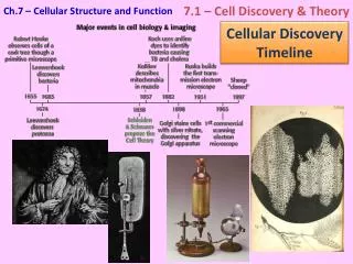
7.1: Cell Discovery and Theory MAIN IDEA: The invention of the microscope led to the discovery of cells.
Types of Microscopes • Light Microscopes • Light waves pass through small organisms, or thin slices of larger ones • Simple – one set of lenses (magnifying glass) • Complex – more than one set of lenses • Magnify up to 1500 times • Dyes often used – kill/distort cells
Types of Microscopes cont’d • Phase contrast microscope • Type of light microscope • Increases contrast – difference btwn light/dark • Cell structures more visible • Can study living cells and processes
Electron Microscopes • Use electron beams instead of light beams • High magnification • View image on screen: micrograph • 2 Types: • Transmission electron microscope (TEM) – electron beam passed through thin slice of specimen; up to 1 million X magnification • Scanning electron microscope (SEM) – views surface of specimen
1 2 3 4 • Light • Phase contrast • TEM • SEM
Early Scientists… • 1665 – Robert Hooke • Also made microscopes • Examined cork – discovered cells • 1600s – Anton von Leeuwenhoek (Dutch lens maker) – made simple microscopes • Observed tiny organisms (bacteria, insect structure, etc.) • Scientists focused only on cell itself
Cell Theory Discovered! • 1700s – 1800s - 2 German biologists: Matthias Schleiden and Theodor Schwann discovered cell theory • Schwann emphasized contents of cells, found similarities between plant and animal cells • 1864 – Louis Pasteur disproved spontaneous generation • By 1880s – scientists could show how cells divide
Cell Theory • All living things are made of one or more cells. • Cells are the units of structure and function in all organisms. • All cells come from pre-existing cells.
Cells – Units of Life • Cell – basic unit of life; in ALL organisms: • Unicellular (one-celled) • Multicellular (many cells, some with special functions) • Complex organisms: cells tissues organs body systems organism
Prokaryotes “Pro-” = early Simple cells No nucleus No membrane-bound organelles Eukaryotes “you” More complex cells Have a nucleus May have other organelles (other membrane-bound cell structures w/specific jobs) 2 Main Cell Types
Prokaryotes • Microscopic: .1 – 10 micrometers – billions in a spoonful of soil, or in your mouth! • Can live in extreme environments • Bacteria • Has DNA, ribosomes, plasma membrane • Some have cell walls, flagella • Divide rapidly • Will not form tissues
Eukaryotes • Larger – about 2-100 micrometers, some even larger (neurons, frog eggs) • Contain membran-bound organelles: • structures for organization • Allow for different reactions to occur in cell at once b/c separate from each other • All have different functions • Cells can form tissues
7.2: The Plasma Membrane MAIN IDEA: A cell’s plasma membrane helps maintain homeostasis.
Plasma Membrane • Thin, flexible boundary between the cell and its environment • Allows nutrients into the cell • Allows waste to leave the cell
Selective Permeability • Ability of plasma membrane to control which substances, and how much of them, enter and leave a cell
Composition of Membrane • Made of phospholipids which form a double layer (phospholipid bilayer)
Fluid Mosaic Model • The phospholipid bilayer allows other molecules to “float” in the membrane. • Proteins, cholesterol, carbohydrates
Proteins • Transmit signals inside the cell • Act as support structures • Provide pathways for substances to enter and leave the cell
Cholesterol • Prevent fatty acid tails from sticking together
Carbohydrates • Identify chemical signals
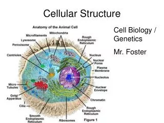
7.3: Cell Structures and Organelles MAIN IDEA: The eukaryotic cell contains organelles.
2 Main Eukaryotic Cell Types • Animal and Plant cells • Many of the same organelles, but some different from each other
Animal Cells Contain centrioles Contain lysosomes NO cell wall NO central vacuole (only small ones) NO chloroplasts Plant Cells NO centrioles NO lysosomes Contain cell wall Contain central vacuole Contain chloroplasts Main Differences
7.4 – Cellular Transport MAIN IDEA: Cellular transport moves substances within a cell, and moves substances into and out of cells.
Passive Transport • Movement of particles across cell membrane without using energy • 3 Types: • Diffusion • Facilitated Diffusion • Osmosis
Diffusion • Movement of particles from an area of high concentration to an area of low concentration
Diffusion Controlled By… • Temperature • Concentration • Pressure • ALL of the above, when increased, increase the amount of collisions particles have with each other, so diffusion occurs more rapidly.
Diffusion and Cells • Diffusion allows for… • Some substances to pass into and out of cells • Substances to spread out within cells • Dynamic Equilibrium • Diffusion into cell = diffusion out of cell • Concentration on either side of membrane is equal, although movement of particles continues
Facilitated Diffusion • Movement of particles across a plasma membrane using proteins • Proteins are specific to certain particles • Channel Proteins – span membrane so particles do not come into contact with nonpolar tails; effective for ion transport • Carrier Proteins – physically bind particles on one side of membrane, and release them on other, changing shape; used for ions, sugars, amino acids
Cellular Structure and Function Facilitated Diffusion Carrier Proteins Channel Proteins
Osmosis • Diffusion of WATER across selectively permeable membrane, from high concentration to low concentration
3 Types of Solutions • Isotonic • Hypotonic • Hypertonic
Isotonic Solution • Water and dissolved substances diffuse into and out of the cell at the same rate.; concentrations equal; cell size, pressure stays the same Blood Cell Plant Cell
Hypotonic Solution • Solute concentration is higher inside the cell than in the solution • Water diffuses into the cell; cell swells • Plant cells don’t burst (b/c of cell wall) Blood Cell Plant Cell
Hypertonic Solution • Solute concentration is higher outside the cell than in the solution • Water diffuses out of the cell (shrinks) Plant Cell Blood Cell
Active Transport • Movement of substances AGAINST the concentration gradient (____ to _____ concentration) • Requires energy • Helps cell maintain homeostasis • Uses carrier proteins called pumps • Some move substances only one way, some move them both ways
Transport of Large Particles • Endocytosis – cell surrounds substance outside of cells, encloses it in a sac/vesicle, and takes it into the cell • Exocytosis – vesicle with a substance is released from the cell