Download

1 / 23
240 likes | 448 Views
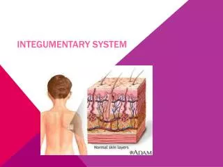
Lab: Integumentary System. Epidermis. Predominant cell type of epidermis are keratinocyes, form stratified squamous epithelium Consists of 5 layers:

E N D
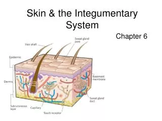
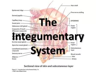
Epidermis • Predominant cell type of epidermis are keratinocyes, form stratified squamous epithelium • Consists of 5 layers: • *Stratum Corneum – largest and most superficial layer, consists of a stratum disjunctum (outer) and stratum conjunctum (inner) layers (as demonstrated on model), several layers of keratinized cells (keratinocytes) that have lost nuclei and organelles • *Stratum lucidum – only present in thick skin (palms of hands and soles of feet), glassy appearance • Stratum granulosum – keratohyaline and lamellated granules • Stratum spinosum – cells have pointy or spiny processes on them • Stratum basale (stratum germinativum) – single row, contains mitotically dividing cells and melanocytes (secrete melanin, giving brown pigmentation to keratinocytes for UV protection)
Epidermis Stratum Spinosum – cells have pointy or spiny processes on them. Stratum Granulosum – contain keratohyaline granules Stratum Lucidum – is present only in thick skin Stratum Cornea – modeled as stratum disjunctum (outer) and stratum conjunctum (inner) . Stratum Basale (Germinativum) – contains dividing cells Melano- cyte Sweat Pore Epidermis and dermis, separated by basement membrane (dark blue) The epithelium is classified as "squamous" based on the cells of the surface layer.
Melanocytes Melanocytes form the skin pigment melanin (can be red, yellow, brown or black in color). found in the stratum basal layer of the epidermis and in the hair follicle. Structure They have long cell processes which extend into the upper layers of the epidermis, and give pigment to the keratinocytes. Thus, a melanocyte supplies 36 keratinocytes on average.
Dermis • Basement Membrane - separates epidermis and dermis • *Dermis • Predominant cell type is fibroblasts which produce collagen, location of arteries, veins and capillaries • Composed of 2 layers: • *Papillary Layer – uppermost layer of dermis, composed primarily of loose CT –areolar, dermal papillae • *Reticular Layer – made of dense irregular CT, 80% of dermis, contains hair follicles, sweat and oil glands (derivatives of the epidermis – below) • *
Dermis Dermal Papillae Papillary Layer – uppermost layer of the dermis, Composed of loose connective tissue, mostly fibroblasts, but also mast cells, macrophages, and leukocytes Reticular Layer – made up of dense irregular connective tissue
Dermal Papillae *Dermal papillae - extend up into epidermal region and contain capillary beds to supply nutrients to surrounding cells *Meissner’s corpuscles – tactile receptors that house sensory structures for touch, commonly found in dermal papillae *Pacinian corpuscles – similar to meissner’s corpuscles, responsible for sensitivity to vibration and pressure
Arterioles (Red) and Venules (Blue) Capillaries Arteries (Red) and Viens (Blue)
Hypodermis • Not technically part of the skin • Loose CT layer, high concentration of adipose tissue
Glands • Sebaceous Glands • Sweat (suderiforous) Glands (2 types) • Apocrine • Eccrine
Glands • Sebaceous Glands – releases an oily secretion (sebum), holocrine mode of secretion (Products are secreted by rupture of gland cells)
Glands • Sweat (suderiforous) Glands (2 types) – Merocrine secretion (products secreted by exocytosis)
Eccrine Sweat Glands • Eccrine – found all over the body and function throughout your life • Abundant on palms, soles and Forehead, • secrete sweat (water, salt, vit C, antibodies, metabolic wastes, etc)
Sweat pore Eccrine gland Sebaceous gland Figure 5.5b
Duct – stratified cuboidalSecretory – simple cuboidal or columnar
Apocrine Sweat Glands • develops during puberty and are most active during adulthood • located in the axillary and anogenital areas (responsible for what we refer to as body odor), • secrete sebum of sweat and fatty substances • Specialized apocrine glands: ceruminous glands (in external ear, secrete cerumen/earwax), mammary glands
Hair • Description – made of hard keratin (dead keratinized cells), derivative of epidermis, extends into dermis • Functions: guard scalp against physical trauma, heat loss and sunlight • Distribution: found all surfaces except palms, soles, lips, nipples, and portions of external genitalia
Hair • Consists of: • Shaft • Root • Follicle • Bulb • Arrector Pili • What is the function of each? What portion produces the hair?