Download
1 / 16
160 likes | 181 Views
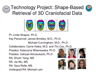
Technology Project: Shape-Based Retrieval of 3D Craniofacial Data. PI: Linda Shapiro, Ph.D. 1 ,2,5 James Brinkley, M.D., Ph.D. 1, 3 ,5 Michael Cunningham, M.D., Ph.D. 3,4, 6 1 Department of Computer Science and Engineering 2 Department of Electrical Engineering

E N D
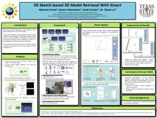
Technology Project: Shape-Based Retrieval of 3D Craniofacial Data PI: Linda Shapiro, Ph.D.1,2,5 James Brinkley, M.D., Ph.D.1,3,5 Michael Cunningham, M.D., Ph.D.3,4,6 1Department of Computer Science and Engineering 2Department of Electrical Engineering 3Department of Biological Structure 4Department of Pediatrics 5Department of Medical Education & Biomedical Informatics 6Craniofacial Center, Seattle Children’s Hospital
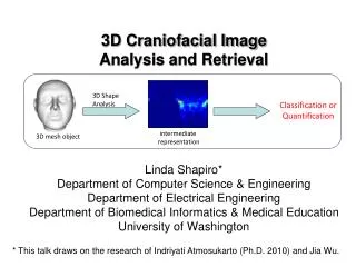
PURPOSE • Researchers at the Seattle Children’s Hospital Craniofacial Center study craniofacial disorders in children. • The goal is to develop principled algorithms to reveal genotype-phenotype disease associations. • This requires the development of computational techniques to represent, quantify, and analyze variants of biological morphology from imaging sources, such as CT scans, MRI scans, and stereo cameras.
Specific Aims • Develop software tools that produce quantitative representations of craniofacial anatomy that can assist in the study of midface hypoplasia and cleft lip and palate. stereo imaging setup 3D mesh two representations of facial shape
Specific Aims 2. Develop tools for quantifying the similarity of craniofacial data between two individuals, between an individual and an average over a selected population, or between two populations. Affected Control New Individual
Specific Aims • Develop mechanisms for organization and retrieval of multimodality 3D craniofacial data based on their quantitative representations.
Specific Aims • Design and implement a prototype system for Craniofacial Information Retrieval (CIR) that incorporates quantification, organization, and retrieval. • Evaluate it on 3D craniofacial data with collaborators from Seattle Children’s Hospital. • Make it available to the FaceBase HUB.
Preliminary Studies - 22q11.2DS • Caused by a microdeletion on chromosome 22 • Multiple different subtle physical manifestations • Midface hypoplasia is one such manifestation Images are for educational purposes only and may not be used in publications.
Shape Quantification - 22q11.2DS • Three different global descriptors with PCA
Preliminary Studies - Deformational Plagiocephaly and Brachycephaly • Caused by persistent pressure • Manifests in flat areas of the back of the head plagiocephaly brachycephaly
Shape Quantification - Plagiocephaly • Shape descriptor: 2D histogram of azimuth and elevation angles of surface normals
Data Sets for Facebase Work • Data from our preliminary studies • 3D mesh data from studies of 22q11.2DS • 189 subjects, 53 affected, 136 controls • 3D mesh data from studies of plagiocephaly and brachycephaly • 462 subjects, 231 referred cases, 231 controls • CT scan data from studies of craniosynostosis • 152 subjects, 99 affected, 53 controls
Prospective Enrollment through FACEBASE: Controls from Dr. Weinberg’s FaceBase proposal (n=3,000) • Retrospective Enrollment From Existing Studies: • Data from clinical care (n*=200) • Data from IRB-approved studies: • 22q11.2DS (n*=100) • Deformational Plagiocephaly (n*=250 cases, 250 controls) • Craniofacial Microsomia (n*=20) • Craniosynostosis (n=250) Data: 3D Image (Mesh only) 3D CT data (if available) Data: 3D image 3D CT data (if available) 3D Data available for analysis in the FaceBase proposal: Shape-Based Retrieval of 3D Craniofacial Data Year 1: ~1000 images Years 2-5: >4000 images Data: 3D image 3D CT data (if available) Data: Full 3D image • Prospective Enrollment (Option Added to Existing SCH Studies): • Data from the following IRB-approved studies: • 22q11.2DS (n=50) • Craniofacial Microsomia (n= 200) • Unilateral cleft lip and palate (n=20) Retrospective Enrollment from the Existing Craniofacial Normative Database Study. Controls (n*=250).
Main Priorities • Software tools that significantly improve the ability to access 3D craniofacial data • Similarity-based retrieval techniques • Craniofacial information retrieval system that can handle both • Multiple modalities of image data • Standard alphanumeric data related to subjects, studies, surveys • Graphical user interface that allows medical users to easily design queries
Hopes for the HUB • A home for both data sets and software tools. • Methodology for sharing data and tools among research groups. • Model for software to be developed. • Forum for discussion among members of the FaceBase Consortium • Application to other species
List of Additional Collaborators • Senior Researchers • Carrie Heike, MD, Seattle Children’s Hospital • Timothy Cox, Ph.D, UW Pediatrics • Postdoc • Katarzyna Wilamowska, Ph.D, Biomedical & Health Informatics (NLM Trainee) • Graduate Students • Indriyati Atmosukarto and Shulin Yang, CSE • Jia Wu and Sara Rolfe, EE