Download
1 / 24
350 likes | 1.22k Views
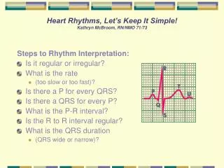
Heart Rhythms, Let’s Keep It Simple! Kathryn McBroom, RN/NMO 71/73. Steps to Rhythm Interpretation: Is it regular or irregular? What is the rate (too slow or too fast)? Is there a P for every QRS? Is there a QRS for every P? What is the P-R interval? Is the R to R interval regular?

E N D
Heart Rhythms, Let’s Keep It Simple!Kathryn McBroom, RN/NMO 71/73 Steps to Rhythm Interpretation: • Is it regular or irregular? • What is the rate • (too slow or too fast)? • Is there a P for every QRS? • Is there a QRS for every P? • What is the P-R interval? • Is the R to R interval regular? • What is the QRS duration • (QRS wide or narrow)?
OK, you have analyzed your strip.When assessing your patient, what do you look for? • Is your patient: • Alert and oriented • Skin warm and dry • Short of breath (at rest or post activity, remember to ask) • Experiencing palpitations (is the pulse slow/fast, regular/irregular). • Complaining of lightheadedness or dizziness Be sure to obtain a set of vital signs
Normal Sinus Rhythm • Originates in the SA node, follows appropriate conduction pathways. • Rhythm: Regular • Rate: 60-100 bpm • Every P has a QRS and every QRS has a P • PRI: .12-.20 seconds • QRS: .8 -.12 seconds, narrow
Sinus Bradycardia • Originates in the SA note. Rate is slower because of sympathetic input or excessive vagal tone. • Seen most often with inferior MI, hypoxia, hypothermia, or drug reactions. • Patient may be asymptomatic. • Rate: < 60 bpm • Every P has a QRS and every QRS has a P • PRI: .12 - .20 seconds • QRS: .8 - .12 seconds, narrow
Sinus Tachycardia • Originates in the SA node. Rapid rhythm which occurs with increased oxygen demand ( exercise, infection, hypovolemia, hypoxia, MI, and to stimulant drugs). • Rhythm: regular/fast • Rate: > 100 bpm • Every P has a QRS and every QRS has a P • PRI: .12 - .20 seconds • QRS: normal
Premature Atrial complex (PAC) • Originates in the atria. Occurs before the normal beat is expected. May occur in the healthy heart. • Can be triggered by anxiety, fever, increased sympathetic input, caffeine and other stimulants, drug interactions, AMI, cardiac ischemia, valvular heart disease, and fever • Rhythm: Irregular with PAC’s • Rate: dependent on rhythm • Every P has a QRS and every QRS has a P • PRI: .12 - .20, may differ from underlying rhythm • QRS: dependent on rhythm
Atrial Fibrillation • Most common cardiac arrhythmia. • May occur with hypertension, ischemia, mitral valve and pericardial disease, MI, aging. • Increased risk for developing atrial thrombus and systemic embolism. Treatment includes anticoagulation, drugs to slow ventricular conduction (rate control) and/or chemical or d/c cardioversion. • Rhythm: irregularly irregular • Rate: slow or fast • No identifiable P’s • QRS usually narrow but may be wide with conduction defect
Atrial Flutter • Characterized by “saw tooth” atrial activity • Conduction ratio to the ventricles 2:1 – 8:1. ( usually 2:1-4:1) • Caused by a reentrant circuit located in the right atrium. • May occur in COPD, hypoxia, intrinsic cardiac disease, valve disease, pericarditis or post operatively. • If >150 bpm, may seriously compromise cardiac output. • Treatment is rate control, cardioversion, surgical or catheter ablation. • Rate: atrial rate 250-400 (generally 300bpm)
Supraventricular Tachycardia/Atrial Tachycardia • There are several different types of SVT/AT, depending on the site of reentry (originates above ventricle) accessory pathway, atrioventricular node, atrium • If this rhythm starts and stops suddenly it is called paroxysmal SVT. • MD may consider vagal maneuvers, antiarrhythmia medications, RFA or surgical modification at site of entry • Rhythm: Regular • Rate: 150-250 bpm • PRI: Dependent on location of “circuit” • QRS: Normal, if accessory pathway used – prolonged (>.12)with delta wave (WPW)
Junctional Rhythm • An escape rhythm serves as a protective mechanism when higher centers in the conduction system fail to fire. • Occurs with increased vagal tone to the sinoatrial node, hypoxemia, and digitalis toxicity • Rhythm: Regular • Rate: 40 – 60 bpm • P wave: • Before QRS, inverted and P-R interval is < .12 • After QRS and usually inverted • Absent • QRS: < .12 seconds, unless prolonged by aberrant conduction
Premature Junctional complex (PJC) • Originates in the atrioventricular junction. • Depending on when the impulse occurs, the P wave may fall before, during or after the QRS complex. • May occur with ischemia, hypoxemia, valvular disease, digitalis. • Rhythm: Irregular with PJC’s • Rate: dependent on rhythm • P wave: • Before QRS, inverted and P-R interval is < .12 • After QRS and usually inverted • Absent • PRI: .12 seconds or less • QRS: < .12 seconds
First Degree AV Block • Occurs when impulses from the atria are consistently delayed during conduction through the AV node. • First degree AV block is a constant and prolonged PR interval. • May result from insult to the AV node, hypoxemia, MI, ischemia, increased vagal tone, aging, beta blockers, calcium channel blockers, digitalis toxicity but is also seen in normal conduction. • Rhythm: Regular • Every P has a QRS and every QRS has a P • PRI: > .20 seconds • QRS < .12
Second Degree AV BlockMobitz I (Wenkebach) • Wenckebach is characterized by progressive delay at the AV node until the impulse is completely blocked. Possible causes are insult to the AV node, hypoxemia, MI, digitalis toxicity, ischemia, and increased vagal tone. This conduction usually does not progress to higher degree heart blocks. • No treatment needed if patient is asymptomatic • Rhythm: Irregular • PRI: progressive lengthening of PRI until dropped beat. (long, longer, drop) • QRS is usually < .12
Second Degree AV Block, Mobitz II • Because the ventricle rate is slow, cardiac output may be decreased • May progress to third degree heart block. • Occurs when some impulses from SA node fail to reach the ventricles • Usually occurs with AMI, degenerative changes in conduction, progressive CAD • Problem usually occurs at the Bundle of HIS or it’s branches • Rhythm is irregular (because of dropped beats) • PRI: remains constant until a block of the AV conduction, resulting is a P wave not being followed by a QRS • Is there a P for every QRS (YES); is there a QRS for every P (NO)? • Treatment: the aim is to improve cardiac output. Consider temporary pacing or permanent pacemaker. Close monitoring and BP support.
Third Degree Heart Block • No conduction through the AV node (“divorced heart”). • Atrial and Ventricular rate and rhythm are independent of one another • Treatment: temp. or permanent pacing • Rhythm is regular (ventricular and atrial, but at diff. rates) • Rate: • Atrial: 60 to 100 • Ventricular 40 to 60 • PRI: will vary with no pattern or regularity • QRS: origin of impulse determines QRS width. • From AV node: QRS will be normal • From Purkinje system: QRS will be wide, rate < 40
Premature Ventricular Contraction • PVC complex may be isolated or occur in pairs or clusters • Primary cause: electrical irritability • Potential for developing dysrhthmias increases in patients with ischemia or progressive heart disease • Treatment: none unless symptomatic • Rhythm: irregular • P wave: usually absent • QRS: greater than .12 seconds and “wide and bizarre”
PVC’s in Couplets • A pattern of two PVC’s following a normal complex. Remember: Three or more PVC’s in a row is VT • A result of ventricular irritability • QRS: > .12 and wide and bizarre • Treatment: close monitoring to assess possibility of ventricular tachycardia, monitor labs (potassium and magnesium)
Ventricular Tachycardia • Three or more beats originating in the ventricle • VT without pulse is treated like ventricular fibrillation (see ACLS algorithm) • Rate usually regular, 100 - 250 bpm • P wave: may be absent, inverted or retrograde • QRS: complexes bizarre, > .12 • Rhythm: may be irregular but usually regular
Idioventricular Rhythm • Escape rhythm (safety mechanism) to prevent ventricular standstill • HIS/purkinje system takes over as the heart’s pacemaker • Treatment: pacing • Rhythm: regular • Rate: 20-40 bpm • P wave: absent • QRS: > .12 seconds (wide and bizarre)
Ventricular Fibrillation • Pt is pulseless i.e. dead • No organized electrical activity • No cardiac output • Causes: MI, ischemia, untreated VT, underlying CAD, acid base imbalance, electrolyte imbalance, hypothermia, • Treatment: ALWAYS immediate unsynchronized defibrillation (see ACLS algorithm). • Rhythm: irregular-coarse or fine, wave form varies in size and shape • No discernible complexes
Pulseless Electrical Activity (PEA) • Electrical activity without pulse (no muscle contraction) • Any rhythm can be seen on monitor, but pt. is pulseless • Causes: MI, hypovolemia, hypoxia, hypothermia, hypo/hyperkalemia, tension pneumothorax, cardiac tamponade, massive pulmonary embolism, acidosis, and drug overdose • Treatment: Identify and correct underlying cause (see ACLS algorithm) • Requires rapid identification and treatment • Rhythm: evidence of organized electrical activity but without pulse • PRI: may appear normal • QRS: may appear normal “Insert any rhythm here” Any rhythm can be seen on the monitor
Asystole • Ventricular standstill, no electrical activity, no cardiac output • Occurs in cardiac arrest, may follow VF or PEA • Remember! No defibrillation with Asystole • Treatment: Epinephrine and Atropine, consider causes (pulmonary embolism, acidosis, tension pneumothorax, hypo/hyperkalemia, hypoxia, cardiac tamponade, hypothermia, drug overdose, MI) • Rate: absent due to absence of ventricular activity. Occassional P wave may be identified. Patient will not have a pulse.
Heart Rhythm Quiz • 1. In Second Degree Type I AV block, the PR interval: a. Varies according to the ventricular response rate b. Progressively lengthens until a QRS complex is dropped. c. Remains constant despite an irregular ventricular rhythm. d. Is un-measurable • 2. You see a rhythm with a narrow complex on the monitor that has a P for QRS and a rate of 165. It is: • Sinus tachycardia • Atrial tachycardia • Atrial fibrillation • Junctional tachycardia • 3. In third degree heart block (CHB) the ventricular response (QRS) is: • Narrow or wide and regular • Wide and irregular • Narrow and irregular • Regular except when a beat is dropped • 4. When 2 or more PVC’s are seen on a strip, it is important to use the following descriptors if applicable: • Couplet/triplet • Multifocal/ Unifocal • Groupings (bigeminy, trigeminy etc.) • All of the above • 5. Characteristics of Normal sinus rhythm are: a. Regular, normal rate with Normal PR and QT intervals b. A P wave for every QRS complex, all P waves are similar in size and shape c. All QRS complexes are similar in size and shape d. All of the above
References • Lippincott Manual of Nursing Practice, eighth edition, pp. 420-429 • Lippincott, Williams and Wilkins, ECG Interpretation, 3rd edition, pp. 65-159 • American Heart Association, ACLS for Healthcare Providers - Algorithm Review. • www.americanheartassociation.com • www.cardiacarrhythmias.com • www.googleimages.com • www.rnceus.com • www.PERD.LLC