Download
1 / 66
670 likes | 989 Views
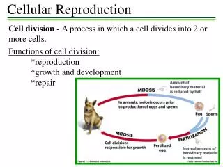
Chapter 14- Cellular reproduction. Where we’re going Review of cell cycle Mostly focusing on regulation of processes Kinases, CDK’s will be critical, along with checkpoints Some review from genetics Mitosis in a bit more depth. What we’ve learned from bacteria (I’ll NOT skip for now .

E N D
Chapter 14- Cellular reproduction Where we’re going • Review of cell cycle • Mostly focusing on regulation of processes • Kinases, CDK’s will be critical, along with checkpoints • Some review from genetics • Mitosis in a bit more depth
What we’ve learned from bacteria (I’ll NOT skip for now • Bacteria grow at a variety of rates, depending upon nutrients available. The cells have to increase to a certain size, and they also have to replicate and partition their DNA. • Initiation of DNA replication is triggered by increased cell size; as the mass increases, DNA replication is initiated. Some cells have multiple initiation events, and the amount of DNA in a cell may vary; • however, the origin:cytoplasm ratio tends to remain constant over a two-fold range.
Once replication has begun, the cell won't divide until it is completed. If you block replication you block cell division: you produce “snakes”. • The models are consistent with the production of a protein that builds up to a certain level in the cell. Initiation then destroys that protein. The cell may also produce a cell division inhibitor, that prevents division until replication and partitioning is complete.
How do we determine these stages? • 1. Generation time: how long does it take to make a new cell? Easily answered by doing a growth curve: seeing how long it takes to double the cell number. • 2. Mitotic index: once you know the generation time, the mitotic index will tell you how long mitosis is: e.g., if 5% of the cells are in mitosis at any one time, and there is a 20 hr generation time, then mitosis lasts 1 hr, approx. (there is a correction factor, because there is always more younger than older cells in a culture)
3. S phase can be calculated similarly; add 3H thymidine briefly, then autoradiograph. The % of cells in S-phase = the length of S phase. • 4. G2-phase can be calculated by labeling briefly, and then determining the earliest time that labeled cells enter mitosis. (14.2)
Important! Fairly short G2 of 3-5 hrs. Essentially a pulse-chase
Models for how cell cycle is controlled- washing machine! Fig. 17-14, MBOC How is the cell cycle like a washing machine? A washer doesn’t start agitating until it’s full of water! It does NOT simply start a few minutes after you hit “start”. Am I big enough?
Cool experiments tell us about the checkpoints • Fuse G1 and S phase cells- G1 goes into S phase. Something is in the S phase cell that will drive the G1 cell into S phase. • Fuse G2 and S phase- nothing! The G2 cell is now resistant to the S phase “factor” • Fuse M phase and other cells- promote condensation
Key players: CYCLINS and CDK’s- Cyclin-dependent protein kinases
Quiz on Friday • Cell Cycle- • How do you tell how long is: M-phase, S phase, G2 • Cyclins? CDK’s?
Ubiquitin added, then into the TUNNEL of DEATH! Active cdc2 kinase actually sets up a positive feedback loop- activating more cdc25, and inactivating wee1. Thus, the amt of active cdc2 kinase EXPLODES!
Some activity, but amplified by active M-CDK M-CDK phosphorylates Wee1, inhibiting the inhibitor! From MBOC, Fig. 17-23
These switches are progressive: After mitosis, there's cell growth; G1 cyclin builds; G1 START; S phase; mitotic cyclin builds---M-phase promoting factor is activated; mitosis.
These move things into Mitosis. Destruction of MPF allows reentry into G1! These move things through S These move the cell through G1-S
Controls to the system There are several controls, such that, even if other conditions are met, the cell won't go on with the cell cycle.
To leave S phase, DNA must be replicated fully, but only once • Stop replication with a poison, and the cell won't enter mitosis. • After S-phase: the re-replication block: cell fusion studies mentioned previously. DNA, once replicated, won't replicate again until after mitosis and the next G1-S transition. • http://www.ncbi.nlm.nih.gov/books/bv.fcgi?rid=mboc4.figgrp.3217 • Fig 17-22 MBOC. There’s a protein that sits on the origin that, in one form, activates the origin, but, after that, it inhibits the origin.
Figure 17-22. The initiation of DNA replication once per cell cycle. The ORC remains associated with a replication origin throughout the cell cycle. In early G1, Cdc6 associates with ORC. Aided by Cdc6, Mcm ring complexes then assemble on the adjacent DNA, resulting in the formation of the pre-replicative complex. The S-Cdk (with assistance from another protein kinase, not shown) then triggers origin firing, assembling DNA polymerase and other replication proteins and activating the Mcm protein rings to migrate along DNA strands as DNA helicases. The S-Cdk also blocks rereplication by causing the dissociation of Cdc6 from origins, its degradation, and the export of all excess Mcm out of the nucleus. Cdc6 and Mcm cannot return to reset an ORC-containing origin for another round of DNA replication until M-Cdk has been inactivated at the end of mitosis .
d. Damaged DNA: p53 is involved in damage repair; cells with damaged DNA repair it before going into mitosis. 14.9. This is a story we’ll want to learn. • The key here is the ATM/ATR proteins- bind to DNA breaks, and becomes ACTIVE. • Once active, it then can phosphorylate Chk2 (if the cell is in G1) or Chk1 (G2- go figure the names!) Chk2-P then phosphorylates p53- the FAMOUS p53- stabilizing it. P53 is a transcription factor- p21 is produced, which inactivates CDK- no movement into S phase. • If the cell is in G2, ATR activates Chk1-P, which phosphorylates Cdc25- the activator of MPF. Once phosphorylated, it’s removed from the nucleus by RAD24- CDK remains inactive, no movement into M phase.
P P Cdc25- key phosphatase for activating CDK
The metaphase-anaphase transition • All the chromosomes have to be attached before anaphase can begin- not just most! • A bit about sister chromosomes- they have securin on them, binding them together preventing anaphase • The APC acts to ubiquitinate securin, marking it for destruction in the tunnel of death (aka proteasome) • MAD2 inhibits APC until all the chromosomes are attached to securin!
APC adds ubiquitin to proteins, destroying key proteins that finish Mitosis. APCCdc20 is what cuts securin, and APCCdh1 is what “cuts” M-CDK SCF Is the ubiquinator during nomal cell growth
Unattached chromosomes have MAD2, which stops APC! When ALL are attached, MAD2 goes away! MAD2 They add ubiquitin, which allows cutting!
Other things • Cell size- probably operates in G1, and may simply be tied to production of cyclin • SEX IN YEAST! When yeast are induced to mate, they can send out a soluble signal that inhibits cell division in nearby yeast until they have mated with the signal producer! Amazing!
Mitosis • Where we’re going- we’ll put a cell biology “spin” on this subject- adding some of the areas that we’ve already covered, mainly from what we’ve learned about the cytoskeleton, to this subject.
The centrosome duplicates the centrioles, MT’s disassemble, then reassemble as spindle fibers
The nucleus disappears- how this happens is not clear. The centromeres assemble kinetochores.
Once attached, the chromosomes head for the middle; there’s much tubulin turnover at the plus end until it reached the equator. VERY strange at the molecular level!
Lots of tubulin flux in the polar and chromosomeal spindle fibers. Treadmilling!
Anaphase A- Chr. Move to opposite poles Anaphase B- poles move farther apart
Anaphase A Anaphase B So the movement is due to new tubulin being added (red)!
Cool experiment that shows that tension’s needed for anaphase!
The furrow is placed mid-way between the nuclei, and is actin-requiring- two experiments