Download
1 / 1
20 likes | 502 Views
Diagnostic dilemma; Cornual Pregnancy Dr Mona Modi, Dr J. Arora, Dr. T. El-Shamy, Ms. S. Sawant. East and North Hertfordshire NHS Trust. Case Report. Discussion. Objectives. Conclusions.

E N D
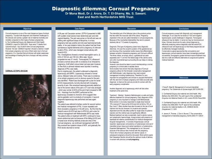
Diagnostic dilemma; Cornual Pregnancy Dr Mona Modi, Dr J. Arora, Dr. T. El-Shamy, Ms. S. Sawant.East and North Hertfordshire NHS Trust. Case Report Discussion Objectives Conclusions Cornual pregnancy is one of the most dangerous types of ectopic pregnancy. It poses both diagnostic and treatment challenges to the clinician and carries a greater risk of maternal morbidity and mortality compared to other types of ectopic pregnancies. In The Confidential Enquiry into Maternal and Child Health (CEMACH ) report for 2000-2002, there were 11 deaths from ruptured ectopic : four of which were Cornual pregnancies. However the last CEMACH reported showed a decline in death from ectopic pregnancy and none of them were due to Cornual pregnancy. It is important that all clinician with better awareness and knowledge of treating this form of ectopic will help continue the trend. CORNUAL ECTOPIC ON SCAN A 38 year old Caucasian woman, G7P3+3 presented to A&E with sudden onset severe lower abdominal pain and mild vaginal bleeding. The pain was worse on the left and was not settling with significant doses of Morphine. A urinary pregnancy test was positive and she was haemodynamically stable. In her past obstetric history the patient had had three spontaneous vaginal deliveries and a pregnancy of unknown location (PUL), which was managed conservatively with serial β-hCG. The investigations showed a normal haemoglobin and a β-hCG of 105 IU/L and 100 IU/L 48hrs later. Serum progesterone was 31 nmol/L. Transvaginal (TV) ultrasound showed a normal uterus with no evidence of an intrauterine pregnancy. A left sided corpus luteal cyst was seen however, no free fluid or adnexal masses were reported. A working diagnosis of PUL was made. Due to ongoing pain, the patient underwent a diagnostic laparoscopy and ERPC. Laparoscopy showed a normal uterus, fallopian tubes and ovaries. There was no evidence of an ectopic pregnancy and no obvious cause for the pain was identified. Patient was discharged with a plan for serial β-hCG measurements, but she returned with increasing severity of pain. She remained haemodynamically stable but due to the severe nature of the pain a CT scan was arranged , which was normal. β-hCG at this point had increased to 468 IU/L . Histology from the ERPC performed at first laparoscopy revealed no chorionic villi to suggest inter-uterine gestation and no products of conception. A second diagnostic laparoscopy was performed and was again negative. The patient was referred to tertiary centre for second opinion and medical management of PUL. A scan repeated over there revealed a pregnancy in the left cornu. By this time the patient’s serum β-hCG had risen to 878 IU/L and a plan for medical management with Methotrexate (MTX) was made. The patient while on treatment with MTX, continued to have severe abdominal pain but because of the falling β-hCG and a repeat scan showing relatively avascular area of the Cornual pregnancy we continued management with MTX. She received two systemic doses of MTX and gradually the HCG levels returned to normal. The interstitial part of the fallopian tube is the proximal portion that lies within the muscular wall of the uterus. Pregnancy implanted in this site are called interstitial or cornual pregnancies. Because of the myometrial distensibility they tend to present relatively late at 7-12 weeks of pregnancy. Diagnosis: This type of pregnancy poses many diagnostic dilemmas. On scan the eccentric position of the gestational sac and thinning of the myometrial mantle means that differentiation between eccentric intrauterine pregnancy and Cornual is difficult. The three criteria: an empty uterus, a gestational sac seen separately and <1 cm from the lateral edge of the uterine cavity and a thin myometrial layer surrounding the sac helps in making the diagnosis. However care should be taken to avoid misinterpreting a normal pregnancy in a bicornuate or septate uterus. Some studies have shown that early diagnosis of Cornual pregnancy allows for first trimester conservative management with Methotrexate. Later diagnosis may need surgical management including hysterectomy. Therefore it is very important for sonographers and clinician to acquaint themselves with this rare form of ectopic pregnancy and make accurate early diagnosis to improve the care we provide for women with this early pregnancy complication Another diagnostic aid is laparoscopy which will also allow treatment at the same time. Treatment: : Medical : Systemic Methotrexate is a safe and highly effective treatment for cornual pregnancy. Early diagnosis of this pregnancy is very important to manage it with Methotrexate. Most women in one study responded to single dose however 15% received 2nd dose as the hCG level did not fall by 15% on Day 4 and 7. Many studies have also focused on the use of local injection into the sac however it is invasive and operator dependent and the side effect profile for single dose of systemic injection and this method was not significantly different. Medical treatment can also occasionally lead to uterine rupture and catastrophic haemorrhage. A large ectopic with presence of heartbeat are relative contraindication for medical management Surgical managementL : It consists of laparoscopic cornual resection, laparoscopic cornuostomy or hysteroscopic removal of interstitial ectopic tissue and radical operations i.e hysterectomy. Expectant Management: This management is not favoured because of the serious risks involved with this pregnancy. Future risk of ectopic pregnancy and uterine rupture in subsequent pregnancy should be discussed. Most authors agree that Caesarean Section should be the optimum mode of delivery in women with previous cornual pregnancy. Cornual pregnancy poses both diagnostic and management challenges. In our case the low levels of hCG and negative laparoscopies posed challenges in diagnosis. Transvaginal ultrasound may be helpful in some but may be inconclusive at times. Clinician should aim for early clinical diagnosis by effectively analyzing various diagnostic aids like serial hCG, ultrasound scan and laparoscopy so that these pregnancies can be effectively managed medically. Conservative surgical approaches like cornuostomy and laparoscopic cornual resection have been increasingly practiced successfully. Sonographic guided minimal invasive treatment may also be a safe and effective alternative to surgical and systemic medical treatment. References 1.Faraj R, Steel M. Management of cornual (interstitial) pregnancy. The Obstetrician & Gynaecologist 2007;9:249-255 2. Confidential Enquiry into maternal and child health. Why Mothers Die 2000-2002. The sixth report of the confidential Enquiries into Maternal Deaths in UK, London:RCOG Press:2004 3. Confidential Enquiry into maternal and child health. Why mothers Die 2006-2008. The 8th report of the confidential enquiries into maternal deaths in UK, CEMACE BJOG (Supplement 1) 1-203 4. Jeremy K, Thomas J, Doo A, Bourne T. The conservative management of interstitial pregnancy. BJOG 2004;111:1283-8