Download
1 / 66
660 likes | 667 Views
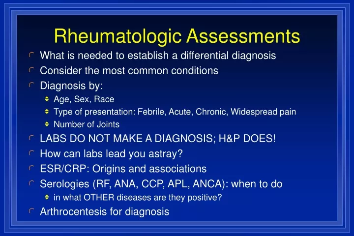
Rheumatologic Assessments. What is needed to establish a differential diagnosis Consider the most common conditions Diagnosis by: Age, Sex, Race Type of presentation: Febrile, Acute, Chronic, Widespread pain Number of Joints LABS DO NOT MAKE A DIAGNOSIS; H&P DOES!

E N D
Rheumatologic Assessments • What is needed to establish a differential diagnosis • Consider the most common conditions • Diagnosis by: • Age, Sex, Race • Type of presentation: Febrile, Acute, Chronic, Widespread pain • Number of Joints • LABS DO NOT MAKE A DIAGNOSIS; H&P DOES! • How can labs lead you astray? • ESR/CRP: Origins and associations • Serologies (RF, ANA, CCP, APL, ANCA): when to do • in what OTHER diseases are they positive? • Arthrocentesis for diagnosis
Common Causes of Joint Pain • Musculoskeletal conditions > 70 million • 315 million MD office visits (Disability 17 million) • Low Back Pain > 5 million per year • Trauma/Fracture • Osteoarthritis 12-20 million • Repetitive strain/injury • Bursitis,Tendinitis;Carpal tunnel syndrome: 2.1 million • Fibromyalgia: 3.7 million • Rheumatoid Arthritis: 2.1-2.5 million • Gout, Pseudogout: 2+ million • Spondyloarthropathy: AS, PsA, Reactive, IBD arthritis (~1.4 mil) • Polymyalgia rheumatica/temporal arteritis • Infectious arthritis
Uncommon Causes of Joint Pain Systemic lupus erythematosus: 239,000 Drug-induced lupus Scleroderma / CREST < 50,000 Mixed Connective Tissue Disease (MCTD) Vasculitis (Polyarteritis nodosa, Wegeners granulomatosus) Inflammatory myositis <50,000 Juvenile arthritis Behcets syndrome Sarcoidosis Relapsing polychrondritis Still’s Disease
Goals of Assessment • Identify “Red Flag” conditions • Conditions with sufficient morbidity/mortality to warrant an expedited diagnosis • Make a timely diagnosis • Common conditions occur commonly • Many MS conditions are self-limiting • Some conditions require serial evaluation over time to make a Dx • Provide relief, reassurance and plan for evaluation and treatment
RED FLAG CONDITIONS • FRACTURE • SEPTIC ARTHRITIS • GOUT/PSEUDOGOUT
Key Questions • Inflammatory vs. Noninflammatory ? • Acute vs. Chronic ? (< or > 6 weeks) • Articular vs. Periarticular ? • Mono/Oligoarthritis vs Polyarthritis ? (Focal) (Widespread) • Are there RED FLAGS?
Monarticular Osteoarthritis Fracture Osteonecrosis Gout or Pseudogout Septic arthritis Lyme disease Reactive arthrtis Tuberculous/Fungal arthritis Sarcoidosis Polyarticular Osteoarthritis Rheumatoid arthritis Psoriatic arthritis Viral arthritis Serum Sickness Juvenile arthritis SLE/PSS/MCTD Mono/Oligo vs Polyarticular
Nonarticular Pain • Fibromyalgia • Fracture • Bursitis, Tendinitis, Enthesitis, Periostitis • Carpal tunnel syndrome • Polymyalgia rheumatica • Sickle Cell Crisis • Raynaud’s phenomenon • Reflex sympathetic dystrophy • Myxedema
Musculoskeletal Complaint • Initial Rheumatic History and Physical Exam to Determine: • 1. Is it articular • 2. Is it acute or chronic? • 3. Is inflammation present? • 4. How many/which joints are involved? • Nonarticular Condition • Trauma/Fracture • Fibromyalgia • Polymyalgia Rheumatica • Bursitis • Tendinitis Is it Articular? No Yes Is Complaint > 6 wks Duration? Yes No • Acute Arthritis • Infectious Arthritis • Gout • Pseudogout • Reiter’s Syndrome • Initial Presentation of • Chronic Arthritis Is Inflammation Present? 1. Is there prolonged morning stiffness? 2. Is there soft tissue swelling? 3. Are there systemic symptoms? 4. Is the ESR or CRP elevated? Acute Chronic No Yes Chronic Inflammatory Arthritis • Chronic Inflammatory • Mono/oligoarthritis • Consider: • Indolent infection • Psoriatic Arthritis • Reiter’s Syndrome • Pauciarticular JA Chronic Noninflammatory Arthritis 1-3 How Many Joints Involved? Are DIP, CMC, Hip or Knee Involved? >3 Chronic Inflammatory Polyarthritis No Yes No • Consider: • Psoriatic Arthritis • Reiter’s Syndrome Is it Symmetric? • Unlikely to be • Osteoarthritis • Consider: • Osteonecrosis • Charcot Arthritis Osteoarthritis Yes • Consider: • SLE • Scleroderma • Polymyositis Rheumatoid Arthritis Are PIP, MCP or MTP Joints Involved? No Yes
Musculoskeletal Complaint < 55 yrs. > 55 yrs.
History: Clues to Diagnosis • Age • Young: JRA, SLE, Reiter's, GC arthritis • Middle: Fibromyalgia, tendinitis, bursitis, LBP RA • Elderly: OA, crystals, PMR, septic, osteoporosis • Sex • Males: Gout, AS, Reiter's syndrome • Females: Fibrositis, RA, SLE, osteoarthritis • Race • White: PMR, GCA and Wegener's • Black: SLE, sarcoidosis • Asian: RA, SLE, Takayasu's arteritis, Behcet's
Onset & Chronology • Acute: Fracture, septic arthritis, gout, rheumatic fever, Reiter's syndrome • Chronic: OA, RA, SLE, psoriatic arthritis, fibromyalgia • Intermittent: gout, pseudogout, Lyme, palindromic rheumatism, Behcet's, Familial Mediterranean Fever • Additive: OA, RA, Reiter's syndrome, psoriatic • Migratory: Viral arthritis (hepatitis B), rheumatic fever, GC arthritis, SLE
Drug – Induced Syndromes • Arthralgias: Quinidine, amphotericin B, cimetadine, quinolones, chronic acyclovir, interferon, IL-2, nicardipine, vaccines • Myalgias/myopathy: Steroids, penicillamine, hydroxychloroquine, AZT, lovastatin, clofibrate, interferon, IL-2, alcohol, cocaine, taxol, colchicine, tryptophan • Gout: Diuretics, ASA, cytotoxics, cyclosporine, alcohol, moonshine, ethambutol • Drug-induced lupus: hydralazine, procainamide, quinidine, methyldopa, INH phenytoin, chlorpromazine, lithium, penicillamine, TCN, TNF inhibitors • Osteopenia: Steroids, chronic heparin, phenytoin, methotrexate • Osteonecrosis: Steroids, alcohol, radiation therapy • Scleroderma/tight skin: Vinyl chloride, bleomycin, pentazocine, solvents, carbidopa, tryptophan, rapeseed oil • Vasculitis: Allopurinol, amphetamines, cocaine, LSD, thiazide, penicillamine, propylthiouracil
Rheumatic Review of Systems • Constitutional: fever, wt loss, fatigue • Ocular: blurred vision, diplopia, conjunctivitis, dry eyes • Oral: dental caries, ulcers, dysphagia, dry mouth • GI: hx ulcers, Abd pain, change in BM, melena, jaundice • Pulm: SOB, DOE, hemoptysis, wheezing • CVS: angina/CP, arrhythmia, HTN, Raynauds • Skin: photosensitivity, alopecia, nails, rash • CNS: HA, Sz, weakness, paraesthesias • Reproductive: sexual dysfunction, promiscuity, genital lesions, miscarriages, impotence • MS: joint pain/swelling, stiffness, ROM/function, nodules
Acute Onset Arthritis • 28 yr. old WF presents with acute onset of knee swelling and pain 7 days ago. Two days later, knee resolved but both wrists began to swell. On day 7, the wrists improved but all PIPs were swollen and tender. • By day 10 she complained of arthritis in PIPs, wrists, knees and ankles. + Tenosynovitis L wrist. AM stiffness was 4 hours. C/O fatigue. Denies fever, rash • She visits her PCP who examines her and orders “Rheumatoscreen Plus” and XRAYs. • He sends her home on OTC ibuprofen, tylenol and Vicks Vapo-Rub.
Acute Onset Arthritis/Rash • Day 14 she returns to PCP with low grade fever, rash (pruritic) on the trunk and extremities. • Exam: symmetric polyarthritis in an RA-like distribution. Tenosynovitis has resolved. Urticarial lesions over trunk and extensor surface of arms. (+)2 cm nontender, left axillary LN. No malar rash, nodules, acne, or Raynauds phenomena. • Investigations?
Acute Onset Migratory Arthritis • WBC = 11.2 • H/H = 13.7 / 38.9 MCV = 89 • ESR = 123 mm/hr • SMA-12 WNL, except albumin = 3.3, AST-67, ALT 77 • ANA negative • RF 31 IU/ml (nl < 30 IU/ ml) • C3 173, C4 28, ASO = 151 Todd units • Uric Acid = 6.6 • Normal SPEP, UPEP, TFT’s, TSH, Ferritin • Others?
Acute Onset Arthritis/Rash • She returns after 1 wk for LN Bx results (negative) • Pt. states her rash and arthritis have nearly resolved. • Exam confirms only mild swelling in knees • However, her sclera are definitely icteric. • Next?
Migratory Arthritis • Viral arthritis (hepatitis B) • Rheumatic fever • Gonococcal arthritis • SLE • Behcets • Hyperlipidemia
Hepatitis B Associated Arthritis • Arthritis and urticaria part of the “prodrome” • Manifestations due to immune complex deposition • Before the Jaundice • Usually while LFTs elevated • Acute onset • Additive (RA like) or migratory (ARF like) arthritis • Often with tenosynovitis • Synovial fluid: inflammatory • Arthritis disappears with onset of Jaundice
Musculoskeletal Exam • Observe patient function (walk, write, turn, rise, etc) • Identify articular vs. periarticular vs. extraarticular • Detailed recording of joint exam (eg, # tender joints) • Specific maneuvers • Tinels sign Median N. Carpal Tunnel syndrome • Finkelsteins ext.pollicis brevisDeQuervains tenosynovitis • Bulge sign Syn.Fluid Suprapatellar pouch Knee effusion • Drop arm sign Complete Rotator Cuff TearTrauma? • McMurray sign Torque on Meniscus Cartilage Tear
RHEUMATOSCREEN PLUS • IgM- RF • ANA • ENA (SSA, SSB, RNP, Sm) • dsDNA-Crithidia • Scl-70, Jo-1 • Histone Abs • Ribosomal P Ab • Coombs • C3, C4 • CH50 • Cryoglobulins • Lupus anticoag. • Cardiolipin Ab • c-ANCA • anti-PR3, -MPO • anti-GBM • SPEP • Lyme titer • HIV • Chlamydia Ab. • Parvovirus B19 • HBV, HCV, HAV • HLA typing • CBC & differential • Chem-20 • Uric acid • Urinalysis • ESR • C-reactive protein • RPR • CPK • Aldolase • ASO • Immune complexs • TFT’s w/ TSH CUSHY LABS INC. “YOUR INDECISION IS OUR BREAD AND BUTTER”
Presbyterian Hosp. CheapoScreen CBC & diff $35.00 Chem-20 $108.00 Urinalysis $30.00 ESR or CRP $25.30 Uric acid $40.00 ANA + RF $ 238.30 CUSHY LABS INC. “YOUR INDECISION IS OUR BREAD AND BUTTER”
Further Investigations • Many conditions are self-limiting • Consider when: • Systemic manifestations (fever, wt.loss, rash, etc) • Trauma (do exam or imaging for Fracture, ligament tear) • Neurologic manifestations • Lack of response to observation & symptomatic Rx (<6wks) • Chronicity ( > 6 weeks)
Common Rheumatic Tests Tests Sensitivity Specificity Rheumatoid 80% 95% Factor Antinuclear 98% 93% Antibody Uric Acid 63% 96%
Acute Phase Reactants • Erythrocyte Sedimentation Rate (nonspecific) • C-Reactive Protein (CRP) • Fibrinogen • Serum Amyloid A (SAA) • Ceruloplasmin • Complement (C3, C4) • Haptoglobin • Ferritin • Other indicators: leukocytosis, thrombocytosis, hypoalbuminemia, anemia of chronic disease
Erythrocyte Sedimentation Rate • ESR : Introduced by Fahraeus 1918 • Mechanisms: Rouleaux formation • Characteristics of RBCs • Shear forces and viscosity of plasma • Bridging forces of macromolecules. High MW fibrinogen tends to lessen the negative charge between RBCs and promotes aggregation. • Methods: Westergren method • Low ESR: Polycythemia, Sickle cell, hemolytic anemia, hemeglobinopathy, spherocytosis, delay, hypofibrinogen, hyperviscosity (Waldenstroms) • High ESR: Anemia, hypercholesterolemia, female, pregnancy, inflammation, malignancy,nephrotic syndrome
ESR & Age M=Age/2 F=Age+10/2
ACP Recommendations for Diagnostic Use of Erythrocyte Sedimentation Rate • The ESR should not be used to screen asymptomatic persons for disease • The ESR should be used selectively and interpreted with caution....Extreme elevation of the ESR seldom occurs in patients with no evidence of serious disease • If there is no immediate explanation for an increased ESR, the physician should repeat the test in several months rather than search for occult disease • The ESR is indicated for the diagnosis and monitoring of temporal arteritis and polymyalgia rheumatica • In diagnosing and monitoring patients with rheumatoid arthritis, the ESR should be used prinicipally to resolve conflicting clinical evidence • The ESR may be helpful in monitoring patients with treated Hodgkin’s disease
Joint Pain and ANA+ • 79 yoWM with schizophrenia and heart failure presents with a 3 month hx of arthralgias affecting the knees, shoulders, elbows. His MD found +ANA 1:80 • He complains of rashes, fever • Says your stethoscope looks like a snake • PMHx: as above, • Meds: Lithium, Thorazine, aldomet, acromycin
Joint Pain and ANA+ • VS: T=98.7 BP=110/75 R=18 P=88 • Alert and oriented x 3 • CVS exam: deferred • Skin: Seborrheic dermatitis • Joints: tender muscles and joints. No synovitis • Labs: W 5.2, H/H 13/39, P178k, ESR 38, neg RF, +ANA 1:80 speckled pattern • Other tests? • Diagnosis?
Reasons for +ANA • Age • Drugs (thorazine, lithium, aldomet, tetracycline) • Drug induced lupus??? • No reason: ANA is low titer nonspecfic pattern • No evidence of SLE
Antinuclear Antibodies • 99.99% of SLE patients are ANA positive • (+) ANA is not diagnostic of SLE • 20 million Americans are ANA+ • 239,000 SLE patients in the USA • Normals 5% ANA+; Elderly ~15% ANA+ • Significance rests w/ Clinical Hx, titer, pattern • Higher the titer, the greater the suspicion of SLE
ANA PATTERN Ag Identified Clinical Correlate Diffuse DeoxyRNP Low titer=Nonspecific Histones Drug-induced lupus Peripheral ds-DNA 50% of SLE (specific) Speckled U1-RNP >90% of MCTD Sm 30% of SLE (specific) Ro (SS-A) Sjogrens 60%, SCLE Neonatal LE, ANA(-)LE La (SS-B) 50% Sjogrens, 15% SLE Scl-70 40% of PSS (diffuse dz) PM-1 PM/DM Jo-1 PM, Lung Dz, Arthritis Nucleolar RNA Polymerase I, others 40% of PSS Centromere Kinetochore 75% CREST (limited dz) Cytoplasmic Ro, ribosomal P SS, SLE psychosis (nonspecific) Cardiolipin Thrombosis,Sp. Abort, Plts AMA, ASMA PBC, Chr. active hepatitis
Antinuclear Antibodies • Virtually present in all SLE patients • Not synonymous with a Dx of SLE • May be present in other conditions: • Drug-induced (procainamide, hydralazine, quinidine, TCN, TNF inhib.) • Age (3X increase > 65 yrs.) • Autoimmune disease • AIHA, Graves, Thyroiditis, RA, PM/DM, Scleroderma, Antiphospholipid syndrome • Chronic Renal or Hepatic disease • Neoplasia associated • Ineffective “screen” for arthritis or lupus • Specificity enhanced when ordered wisely
Frequency in SLE Autoantibody Frequency • dsDNA 30-70% • Sm 20-40% • RNP 40-60% • Ro 10-15% • Ribosomal P 5-10% • Histones 30% • ACA 40-50% Egner W, J Clin Pathol 53:424, 2000
ANA Associations Sensitivity (%) Condition Lane Kavanaugh • Drug-induced LE 100 • SLE 99 95-100 • Scleroderma 97 60-80 • Sjogrens 96 40-70 • MCTD 93 ~100 • PM/DM 78 30-80 • RA 40 30-50 • Vasculitis 15 • JRA 20-50 • Raynauds 20-60 Lane SK, Gravel JW, Am Fam Phys 65:1073, 2002 Kavanaugh AF, et al; Arch Pathol Lab Med 124: 71, 2000