Download
1 / 28
280 likes | 449 Views
Psoriasis & Skin Cancer. Revision. Dermatology History. PC What’s the problem? Where is it? How long has it been there? Hx Of PC What did it look like to begin with? Has it changed? If so, how? Itchy? Painful? Bleeding? Aggravating/ relieving factors

E N D
Psoriasis & Skin Cancer Revision
Dermatology History PC What’s the problem? Where is it? How long has it been there? Hx Of PC What did it look like to begin with? Has it changed? If so, how? Itchy? Painful? Bleeding? Aggravating/ relieving factors Previous & current treatments (effective or not) Recent contact with diseases? Stressful events? Illness? Travel? History of sunburn, use of tanning machines Skin type? (1-4)
Past medical history • History of atopy i.e. asthma, allergic rhinitis, eczema • History of skin cancer & any suspicious skin lesions • Medication & allergies Family history Social history Occupation- alcohol gel, over washing of hands, gloves Improvement of lesions when on annual leave Smoke/Drink? ICE
Examination • INSPECT • DESCRIBE • PALPATE • SYSTEMATIC CHECK
INSPECT • Where are the lesions & how many are there? • Is there a pattern i.e extensors affected only DESCRIBESCAM • Size (at the widest diameter) & Shape • Colour • Associated secondary change • Margin (border)
If lesion is pigmented ABCDE • Asymmetry • Irregular Border • Two or more Colours within the lesion • Diameter > 6mm • Evolution- change in size, shape over time? Started to bleed?
PALPATE & comment on: • Consistency • Mobility • Tenderness • Temperature • SYSTEMATIC CHECK Examine: • Nails • Scalp • Hair • mucous membranes • Regional lymph nodes • General examination
Psoriasis 2 2 1 3 3 1 Describe the lesion 2 What is this? 3 What is this?
Psoriasis Chronic inflammatory skin disease “Hyperproliferation of keratinocytes & inflammatory cell infiltration” Chronic plaque psoriasis most common type Other types include: • Guttate (raindrop lesions), • Seborrhoeic (naso-labial and retro-auricular), • Flexural (body folds), • Pustular (palmar-plantar) • Erythrodermic (total body redness)
Which type? Seborrhoeic Pustular Erythrodermic Flexural Guttate
2% of population in UK Complex interaction of genetic, immunological &environmental factors Precipitating factors inc: -Trauma -Infection (e.g. Strep throat) -Drugs -Stress -Alcohol
Well-demarcated, erythematous, scaly PLAQUES Extensor surfaces of the body & over scalp Auspitz sign (Gentle removal of scales = capillary bleeding) 50% have nail changes (e.g. pitting, onycholysis) 5-8% assc. psoriatic arthropathy: • Symmetrical polyarthritis • Asymmetrical oligomonoarthritis • Lone distal interphalangeal disease • Psoriatic spondylosis • Arthritis mutilans Lesions itchy, burning, painful
Management Avoid precipitating factors, emollientsto reduce scales • Topical therapies (localised & mild psoriasis): -Vitamin D analogues i.e calcitrol -Topical corticosteroids -Coal tar preparations inc. scalp treatments -Dithranol -Topical retinoids i.e Tarazotene -Keratolytics (Urea based creams)
Phototherapy (extensive disease) • - Mainly UVA • -UVB can be used when UVA fails- can cause sunburn • Oral therapies (extensive, severe psoriasis & psoriasis with systemic involvement) -Methotrexate -Retinoids -Ciclosporin -Mycophenolate mofetil -Biological agents (e.g. infliximab, etanercept, efalizumab)
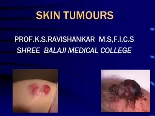
Skin Cancer Skin Cancer Skin Cancer SCC BCC Solar Keratosis Malignant Melanoma
Skin cancer can be divided into: • Non-melanoma (basal cell carcinoma & squamous cell carcinoma) • Melanoma (malignant melanoma).
Basal Cell Carcinoma Slow-growing, locally invasivetumour epidermal keratinocytes- affect basal layers of skin Malignant but rarely metastasises Most common malignant skin tumour Risk factors include: -UV exposure -History of frequent/ severe sunburn in childhood -Skin type 1 -Increasing age -Being male -Immunosuppression -Previous history of skin cancer -Genetic predisposition
Nodular most common • Small, skin-coloured papule or nodule • Surface telangiectasia, • Pearly rolled edge • May have necrotic/ ulcerated centre rodent ulcer • Head & neck involvement • Management • Surgical excision • Radiotherapy - when surgery is not appropriate • Cryotherapy • Topical photodynamic therapy • Topical treatment (imiquimod cream) -small, low-risk lesions
Squamous Cell Carcinoma Locally invasive malignant tumour of epidermal keratinocytes - affect squamous layer of skin. Potential to metastasise Causes Risk factors include: • Excessive UV exposure • Actinic/ Solar keratoses • chronic inflammation (leg ulcers, wound scars) • Immunosuppression • Genetic predisposition
Keratotic (scaly, crusty), ill-defined nodule • May ulcerate • Management • Surgical excision - treatment of choice • Radiotherapy - for large, non-resectable tumours
Malignant Melanoma Invasive malignant tumour of epidermal melanocytes Potential to metastasise Causes Risk factors include: • Excessive UV exposure • Skin type 1 • History of multiple moles/ atypical moles • Family history or previous history of melanoma The ‘ABCDE Symptoms’ rule → What are they? Legs in women & trunk in men
Superficial Spreading Melanoma Nodular melanoma Name the types of melanoma Acral Lentiginous Melanoma Lentigo Maligna Melanoma
Types: • Superficial spreading melanoma – lower limbs, young & middle-aged, intermittent high intensity UV exposure • Nodular melanoma - trunk, in young & middle-aged adults, intermittent high-intensity UV exposure • Lentigo maligna melanoma - face, elderly pop, long-term cumulative UV exposure • Acral lentiginous melanoma - palms, soles & nail beds, elderly pop, no clear relation with UV exposure
Management • Surgical excision • Radiotherapy • Chemotherapy for mets
Prognosis Recurrence based on Breslow thickness <0.76mm thick – low risk 0.76mm-1.5mm thick – medium risk >1.5mm thick – high risk 5-year survival rates based on TNM classification Stage 1 (T <2mm thick, N0, M0) – 90% Stage 2 (T>2mm thick, N0, M0) – 80% Stage 3 (N≥1, M0) – 40- 50% Stage 4 (M ≥ 1) – 20-30%
Thank You For Listening & Good Luck