Download
1 / 24
240 likes | 297 Views
Exercise 2:. 2A: Microscopy. Announcements. Post Lab 2 is assigned today and due by the time your lab meets next. Pre Lab 3 will be available on Wednesday at 5 PM and is also due by the time your lab meets next. LNA Bacteria is assigned today, and due by the time your lab meets next*.

E N D
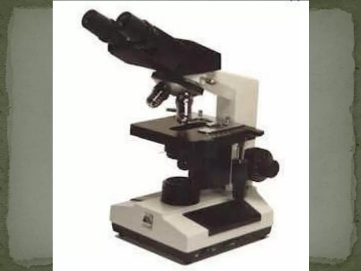
Exercise 2: 2A: Microscopy
Announcements • Post Lab 2 is assigned today and due by the time your lab meets next. • Pre Lab 3 will be available on Wednesday at 5 PM and is also due by the time your lab meets next. • LNA Bacteria is assigned today, and due by the time your lab meets next*. • Pre-Lab Write for LNA 3 is due within the first 5 minutes of lab next week.
Goals • Develop working knowledge of a brightfield light microscope • Discern between different types of microscopy • Practice techniques: objectives, oil, wet mount, measurements
Practice • Practice with the objectives, focusing, and positioning using the prepared slides. • Use your lab manual as a reference if you are having trouble pages 29-30.
Observation of Bacteria • Sphere • cocci • Rod • bacilli • Spiral • Spirochete and spirilla
Exercise 2: 2B: Bacteria
Goals • Become familiar with the scientific process by generating hypotheses, making predictions and designing experiments • Determine potential sources of microbial contamination in the laboratory • Obtain a pure culture of bacteria by streaking for isolated colonies on solid media
Bacteria • Prokaryotes: lack a nuclear membrane • Small, single cell organisms • Exist in huge numbers in small amounts of material • Found almost everywhere
Contamination • Look for contamination by testing for growth on bacterial medium.
Pure Cultures • A population of cells, all of which are descended from a single cell.
Growth Media • Liquid or Solid • Agar • Non-toxic • Remains solid at high temperatures • Not used as a nutrient • Defined or Complex
Sterilization • To kill all living organisms • Autoclaving • Baking • Alcohol • Flaming • Filtration
Aseptic Technique • Inoculating Loop • Serological Pipettes • Microliter Pipettes
Colony • Cluster of cells visible to the naked eye • Facilitating: • Isolation of pure cultures • Enumeration of cell concentration in liquid suspensions • ID of bacterial species based upon the appearance of the colonies
Spreading Plates • A diluted suspension is pipetted directly onto the surface of an agar plate and spread across the surface using a sterile glass spreader.
Determining the number of viable bacteria • The number of colonies on a plate is assumed to be equal to the number of viable cells which were spread on the plate • Limiting Factors • No more than 0.2 ml of cell suspension should be spread on the plate • Resulting in 30 to 300 cells spread on the plate
Determining Cell Mass by Measuring Turbidity • The number of cells per ml is directly proportional to the mass of cells per ml • Using Spectrophotometers to measure Optical Density (OD) • Light is lost as it passes through the suspension because it is scattered and absorbed by the cells. • Data can be used to construct a standard curve.
Bacterial Stains • Positive Stains: cells pick up color • Negative Stains: background color, cells appear white
Procedure Part I • Labeling plates • Label the bottom of the agar plate • Write on the periphery of the plate • Write your name, section, date, and location • Use the sterile swab to collect your contaminant and put it on the agar plate • Incubate plates for 48 hours at 37ºC
Procedure Part II • Isolate Pure Cultures • Using Aseptic Technique • E. coli • Use Streak Techniques
First LNA: Due Next Lab • You should come to lab next with with your lab notebook assignment. • There will be enough time at the beginning of next lab to observe your plates and finish the results and conclusions. • Also remember to have your Pre-Lab write-up (purpose, procedure, data table) for Exercise 3: Enzymes at the beginning of class.