Download
1 / 15
180 likes | 491 Views
Biomedical Image Analysis Rangaraj M. Rangayyan. Ch. 6 Analysis of Shape Sections 6.3-6.8. Presentation March 10th 2005 Jukka Parviainen Yevhen Hlushchuk. Outline. filtering – Fourier descriptors (Sec. 6.3) convex and concave contours (Sec. 6.4-6.5) a few scalar shape descriptors

E N D
Biomedical Image AnalysisRangaraj M. Rangayyan Ch. 6 Analysis of Shape Sections 6.3-6.8 Presentation March 10th 2005 Jukka Parviainen Yevhen Hlushchuk
Outline • filtering – Fourier descriptors (Sec. 6.3) • convex and concave contours (Sec. 6.4-6.5) • a few scalar shape descriptors • applications • summary & discussion T-61.182 Parviainen, Hlushchuk
Books at table • Sonka, Hlavac, Boyle: ”Image processing, analysis and machine vision” • chapter 6: Shape representation and description • Gonzalez, Woods: ”Digital image processing” • chapter 11: Representation and description • (1/N)-multiplier in kD-DFT/IDFT • division to boundary-based and region-based • Rangayyaan focuses on boundary-based • chapters 7 & 8 about textures and patterns T-61.182 Parviainen, Hlushchuk
Introduction • continuation from segmentation process • tools and features • chain coding • signatures, inflections (segmentation of contours), skeletons • compactness (cf), moments (mf), chord-length statistics T-61.182 Parviainen, Hlushchuk
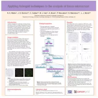
Applications in mammograms • applications rely on contour-based shapes • benign mass contours are often convex, smooth, oval ”simple”, see Fig. 6.9 • malignant tumors typically concave, spicular ”complex”, see Fig. 6.8 T-61.182 Parviainen, Hlushchuk
Benign and malignant masses Fig. 6.8 ”complex” Fig. 6.9 ”simple” T-61.182 Parviainen, Hlushchuk
Fourier descriptors (1), Sec. 6.3 • contour with N points having coordinates {x(n), y(n)}, n = 0 ... N-1 • now z(n) = x(n) + j y(n) 2D to 1D • consider 1D DFT/IDFT Z(k) of complex z(n) • error in (6.55)-(6.56): only one (1/N) • DC-component Z(0) (mean value) • idea: take only K components (K < N) • high frequency components not needed T-61.182 Parviainen, Hlushchuk
Fourier descriptors (2) • example: a square contour (Fig. 11.14/G) • N = 64 points • K = 2 .. 62 components • small K usually enough • square (non-differentials) hard with Fourier kernel T-61.182 Parviainen, Hlushchuk
Fourier descriptors (3) • all Fourier transformations can be applied (Table 11.1) • a scalar shape factor (ff) using norm. F-descriptors • ff is invariant to translation, rotation, starting point, and contour size • ff increases as object shape becomes more complex T-61.182 Parviainen, Hlushchuk
concavity = koveruus Fractional Concavity (Sec. 6.4) • fractional concavity (f_cc) • segmentation of contour with inflection poins: convex part concave part • cumulative length of concave segments • close to 0 when ”simple” region, close to 1 when ”complex” region • Fig. 6.8 f_cc = 0.47, and Fig. 6.9 f_cc = 0.16 T-61.182 Parviainen, Hlushchuk
spicularity = piikkimäisyys Spicularity (Sec. 6.5) • spiculation index (SI): M segments s_m between inflection poins and corresponding angles th_m • Eq. 6.59: if th is 90 degress cos(th)=0 • example in Fig. 6.23 T-61.182 Parviainen, Hlushchuk
Practical problems • Rangayyan: in real life edges are very difficult to detect either manually or by computer • open boundaries • overlapping classes • geometric distortions 3D 2D • texture and gradient features in next chapters T-61.182 Parviainen, Hlushchuk
Applications in mammograms • applications rely on contour-based shapes • benign mass contours are often smooth, oval, convex • malignant tumors typically concave, spicular • studies with shape factors • Sec 6.6: mf, ff, cf • Sec 6.7: cf, f_cc, SI • benign and malignant classes overlapping T-61.182 Parviainen, Hlushchuk
Summary & discussion • several features from signatures to shape descriptors • measurement in scale ”simple” ”complex” • computation of features requires good accuracy with boundaries • otherwise results not reliable T-61.182 Parviainen, Hlushchuk
Matlab Image Processing Toolbox • help images • version Matlab 5.3 - 7, IPT 2.2 - 4 • help iptdemos T-61.182 Parviainen, Hlushchuk