Download
1 / 30
370 likes | 2.05k Views
Scaphoid fractures. Dr Jaycen Cruickshank MBBS FACEM MCR Director of Emergency Medicine, Ballarat Health Services, Ballarat, VIC, AUSTRALIA Senior Lecturer in Emergency Medicine Rural Clinical School Ballarat, School of Rural Health, School of Medicine, University of Melbourne.

E N D
Scaphoid fractures Dr Jaycen Cruickshank MBBS FACEM MCR Director of Emergency Medicine, Ballarat Health Services, Ballarat, VIC, AUSTRALIA Senior Lecturer in Emergency Medicine Rural Clinical School Ballarat, School of Rural Health, School of Medicine, University of Melbourne
Scaphoid fractures Diagnosis Don’t miss them, other fractures when using advanced imaging Guidelines Not widely used, junior staff could use a consistent approach No Australian guideline. Management “clinical scaphoid fracture” Confirmed - operative vs non operative, more details? www.scaphoidfracture.com.au One attempt to insert something humerus
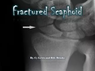
Scaphoid Fractures • Common • High frequency of complications, • this increases when the diagnosis is delayed. • Non-union, delayed union, osteonecrosis and delayed osteoarthritis have been shown to result from scaphoid fractures, with the chance of complications increasing with delayed diagnosis (Langhoff and Anderson 1988). • Mechanism of injury = fall on outstretched hand. • Clinical sign = tenderness - anatomic snuffbox.
CLINICAL EXAMINATION AND X-RAY • Clinical examination is not specific as most injuries that result in joint effusion produce snuffbox tenderness. • Anatomical snuff box • Axial compression of thumb • AP compression scaphoid • X-ray good, but not perfect • Leslie and Dickson reported that 98% of fractures were visible on initial x-ray in their study of 222 confirmed scaphoid fractures, however this number has been reported to be as low as 75-80%. • MRI and CT both demonstrate fractures when the initial x-ray was normal.
WHAT NEXT? • Patients who have a normal x-ray but still have clinical suspicion of fracture are defined as having a “clinical scaphoid fracture”. • Historically these patients are treated with plaster cast immobilization and day 10 review, repeat imaging • Still common, especially in kids. • Recent studies have advocated the use of early advanced medical imaging to limit the time spent in plaster, which affects both patient and community. • MRI • Bone scan • CT • Ultrasound
MRI • MRI has proven to be good for the early diagnosis of scaphoid fracture. Several studies have confirmed that it provides reliable results, and as such have advocated its use. • The American Medical Association list MRI as the gold standard for scaphoid fracture diagnosis. • In Australia: • MRI is expensive (MBS $440) and is difficult to obtain, and a specialist provider number is required for medicare rebate.
MRI - critical evaluation Demonstrated accurate diagnosis of scaphoid and other nearby fractures, with reported 100% negative predictive value, sensitivity and specificity. MRI very reliable (precise) with kappa values of 0.8-0.95. MRI is very sensitive at detecting bone marrow oedema. It is now well documented that patients with clinical scaphoid fracture, have not only scaphoid fractures but other fractures demonstrated on MRI. The prevalence of scaphoid fracture ranges from 13-19%, and other fractures collectively from19% to 24%. This leaves approximately two thirds of patients with no demonstrable fracture. • Mitchell DG, Kressel HY. MR imaging of early avascular necrosis. Radiology. 1988; 169: 281-2. • Cruickshank J, Meakin A, Breadmore R, et al. Early computerized tomography accurately determines the presence or absence of scaphoid and other fractures. Emerg Med Australas. 2007; 19: 223-8. • Kumar S, O'Connor A, Despois M, Galloway H. Use of early magnetic resonance imaging in the diagnosis of occult scaphoid fractures: the CAST Study (Canberra Area Scaphoid Trial). The New Zealand medical journal. 2005; 118: U1296. • Murphy DG, Eisenhauer MA, Powe J, Pavlofsky W. Can a day 4 bone scan accurately determine the presence or absence of scaphoid fracture? Annals of emergency medicine. 1995; 26: 434-8. • Beeres FJ, Hogervorst M, den Hollander P, Rhemrev S. Outcome of routine bone scintigraphy in suspected scaphoid fractures. Injury. 2005; 36: 1233-6. • Biondetti PR, Vannier MW, Gilula LA, Knapp RH. Three-dimensional surface reconstruction of the carpal bones from CT scans: transaxial versus coronal technique. Comput Med Imaging Graph. 1988; 12: 67-73. • Jonsson K, Jonsson A, Sloth M, Kopylov P, Wingstrand H. CT of the wrist in suspected scaphoid fracture. Acta Radiol. 1992; 33: 500-1. • Roolker W, Tiel-van Buul MM, Ritt MJ, Verbeeten B, Jr., Griffioen FM, Broekhuizen AH. Experimental evaluation of scaphoid X-series, carpal box radiographs, planar tomography, computed tomography, and magnetic resonance imaging in the diagnosis of scaphoid fracture. The Journal of trauma. 1997; 42: 247-53.
More critical… • The significance of the MRI finding of bone marrow oedema, a bone bruise: without fracture, following trauma to the scaphoid has been debated, with recent evidence that it is a benign injury and is unlikely to result in long-term morbidity in the form of non-union. • A definition of fracture has normally been a disruption of the cortex (edge) or trabecular pattern (within the bone). There is evidence to suggest that MRI is superior in detecting trabecular fractures than CT, but CT is superior in detecting cortical fractures. • Kappa is only reported between pairs of observers for MRI. • When advanced medical imaging depicts fractures not evident on the existing reference standard, it is inappropriate for authors to suggest that bone scan is prone to false positives when it suggests a fracture that is not evident on delayed x-rays, but to then declare that MRI detects fractures not evident on plain x-ray and is thus more accurate than delayed x-rays. • La Hei N, McFadyen I, Brock M, Field J. Scaphoid bone bruising--probably not the precursor of asymptomatic non-union of the scaphoid. The Journal of hand surgery, European volume. 2007; 32: 337-40. • Raby N. Magnetic resonance imaging of suspected scaphoid fractures using a low field dedicated extremity MR system. Clinical radiology. 2001; 56: • Robinson P. Gold--now you see it, now you don't. Br J Radiol. 2003; 76: 923-.
For scaphoid fracture, gold standards…like Australian Political Partiessample sizes, <100.
BONE SCAN • Bone scans have also shown to aid the diagnosis of scaphoid fracture at an early stage, Day 4. • However, it has been reported that bone scan has a false-positive rate of up to 25% when compared to delayed x-ray. • Bone scan (MBS $ 300) also involves a high radiation dose compared to CT (4.6mSV compared to 0.5mSV).
CT • Several small studies have advocated the use of CT (MBS $220) in the diagnosis of scaphoid fracture. Sensitivity and specificity have been reported to be as high as 100%.
Guidelines for evaluation of wrist injuries • ? impact of such guidelines on the quality of care, patient outcomes, and patient satisfaction.
53 patients with normal CT Time off work mean =1.6 days Plaster mean = 2.7 days MRI if ongoing pain confirmed no fractures missed. Satisfied patients mean 4.2/5 score. 28 fractures, 25 patients 6 scaphoids * 5 triquetral * 4 radius 2 lunate 2 trapezium 2 trapezoid 3 metacarpals (1st 2nd 3rd ) 1 capitate and hamate * one with lunate Results
INTEROBSERVER RELIABILITY -is the gold standard precise, repeatable?What the radiology journals do not want to publish.
Literature – interobserver reliablity MRI Interobserver reliability has been reported as near perfect (k >0.8, and k = 0.95). BONE SCAN Interobserver reliability has been reported as excellent for static phase bone scans (k = 0.81) CT Interobserver reliability has been reported as high (k = 0.76) between different specialties, and excellent (k = 0.86) between two radiologists.
Our study (intraobserver reliability of CT in clinical scaphoid fracture) • 9 radiologists report 15 CT scans each, a sample size of 135. • Sample - stratified randomisation. - scaphoids, others, normals. • kappa value = 0.88 (95% CI = 0.80 – 0.96) scaphoid fracture • 0.56 (95% CI = 0.48 – 0.64) for any fracture. • One radiologist diagnosing twice as many fractures as the rest… • K = 0.93… and 0.7 • Extrapolate… ? • Interobserver reliability for CT between nine observers similar to MRI, between two observers.
Guidelines • National guidelines would be good • Local implementation is required • Implementation of a change in practise in a research setting allowed strict adherence to pathway, patient consent, evaluation of a number of outcomes. • www.scaphoidfracture.com.au
Conclusions • CT has excellent interobserver reliability for diagnosis of scaphoid fracture, comparable to MRI. • CT may have an important role to play in the clinical pathway leading to diagnosis of scaphoid fracture, but clinicians and patients need to be aware of the limitations. • CT is expensive compared to plain radiographs, • and there is a risk of false-positive diagnoses. • This particularly applies to fractures other than the scaphoid.
Management of scaphoid fractures Colles cast versus scaphoid cast: One trial only compared Colles cast to scaphoid cast (Clay 1991). The trial investigated 291 patients, 148 in the Colles cast group and 143 in the Scaphoid group. The main outcome was the union rate. The union was diagnosed on clinical and radiological bases (plain X-ray only). There has been no significant difference between the two treatment groups (Odds ratio 0.96 [0.45-2.07], p-value 0.92).
Operative vs. non-operative treatment Seven trials (Bond 2001; Arora 2007; Dias 2008, McQueen 2008; Adolfsson 2001; Saeden 2001; Vinnars 2008). Studied outcomes included union rate, time to union, ROM, Grip strength, complications and cost. Pooled data - higher union rate in the operative group (Odds ratio 2.81[1.13-6.96]; p-value 0.03). Higher rate of complications in the operative group (Odds ratio 4.20 [2.33-7.65]; p-value 0.0001). Subgroup analysis showed that there was no significant difference in the union rate and complication rate in trials that used percutaneous techniques. In contrast to open technique, there was significant difference in the union rate as well as complications. The ROM, grip strength and return to work data can not be pooled because they have been reported in variable ways. Cautious analysis of the result shows that there is no substantive difference in the ROM, but there is a consistent trend that operation may improve strength and early return to work. However, this remains to be proven. Two trials provided data about the cost effectiveness of operative treatment versus non operative treatment (Arora 2007 and Vinnars 2008). As expected the data was non parametric and could not be pooled. Non operative treatment cost is relatively similar in both trials (2363 Euros and 2507 Euros respectively), but the operative cost is surprisingly low in Arora's study (2097 Euros and 3155 Euros).
Now. The future • Don’t miss the diagnosis • Local guidelines • Future research; better, bigger. • Multi-centre research • Diagnosis • Management • Patient outcomes • Good summary: http://www.springerlink.com/content/q2166j5927231670