Download
1 / 39
430 likes | 642 Views
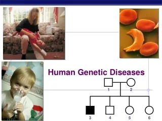
Genetic Diseases. All human diseases can be divided into three categories: - genetically determined - environmentally determined - combination of both (both genetic and environmental factors play a role)

E N D
All human diseases can be divided into three categories: - genetically determined - environmentally determined - combination of both (both genetic and environmental factors play a role) Rapid and continuing progress in molecular research have revealed genetic component in many so-called environmental diseases (e.g. susceptibility to bacterial infections or immune response to them can be influenced by genetic factors) Commonly used adjectives - hereditary = derived from one´s parent - familial = transmitted through generations and affecting several members of a family - congenital = present at birth not all genetic disorders are congenital (Huntington disease: 3rd-4th decade) not all congenital diseases are of genetic origin (congenital syphylis, toxoplasmosis)
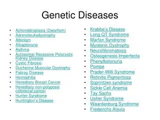
Major categories of genetic diseases • Mendelian disorders • single-gene mutation of large effect • - uncommon conditions (storage diseases, inborn errors of metabolism) • - usually hereditary and familial • Diseases with multifactorial (polygenic) inheritance • - defects of multiple genes with small effect + environmental influences • - some very common diseases (arterial hypertension, diabetes mellitus) • Cytogenetic disorders • - numeric or structural abnormalities of chromosomes
Mendelian disorders more than 5,000 disorders, many of them rare although individually rare, together they account for 1% of adult hospital admissions and 6-8% of pediatric hospital admissions Patterns of inheritance - autosomal dominant - autosomal recessive - X-linked codominance: both alleles of a gene pair fully expressed in the heterozygote (e.g. blood group antigens) polymorphism: presence of many allelic forms of a single gene pleiotropy: single-gene mutation may lead to many phenotypic effects, e.g. Marfan syndrome: widespread effects affecting skeleton, eye and cardiovascular system genetic heterogeneity: several different types of mutation can cause the same phenotypic effect, e.g. retinitis pigmentosa (abnormal retinal pigmentation leading to visual impairment) can be caused by several different mutations
Autosomal dominant disorders - manifested in heterozygous state (only one allele mutant) - at least one parent is affected - new mutation: both parents of affected person are healthy - when affected person marries unafected one → every child 50% chance of having the disease - both males and females can be affected - both males and females can transmit the disease clinical symptoms: 50% reduction in normal gene product - structure proteins and receptors involved - enzyme proteins usually not affected (50% loss of enzyme activity can be compensated for) clinical presentation can be modified by: reduced penetrance: some persons carrying mutant gene are phenotypically normal variable expressivity: all persons carrying mutant gene affected but in different extent among them, e.g. NF1 (from brownish skin pigmentations to multiple tumors and skeletal deformities)
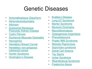
Common autosomal dominant disorders Nervous Huntington disease neurofibromatosis 1 and 2 myotonic dystrophy tuberous sclerosis Urinary polycystic kidney disease (adult type) Hematopoietic hereditary spherocytosis von Willebrand disease GIT familial polyposis coli Skeletal Marfan syndrom Ehlers-Danlos syndrome (some variants) osteogenesis imperfecta achondroplasia Metabolic familial hypercholesterolemia
Marfan syndrome • - abnormality of fibrillin 1 (glycoprotein component of elastic fibers) • - encoded by FBN1 gene (15q21), more than 500 mutations found • - prevalence 1 in 20,000 • - 75% cases familial • - connective tissue throughout the body affected • principal clinical manifestations related to skeleton, eye and cardiovascular system • Skeletal abnormalities (overgrowth of bones) • - slender and elongated habitus, dolichostenomelia (abnormally long legs and arms), arachnodactyly (abnormally long fingers) • - high-arched (gothic) palate • - hyperextensibility of joints • - spinal deformities (kyphoscoliosis) • - chest deformity (depressed sternum - pectus excavatum, pigeon breast)
Ocular changes - bilateral dislocation of lens (weakness of its suspensory apparatus; ciliary zonules are made up exclusively of fibrilin) Abnormalities of cardiovascular system - most serious - fragmentation of elastic fibers of tunica media - aneurysmal dilation of aorta → rupture (most common cause of death) - aortic dissection - dilation of aortic valve ring (loss of medial support) → aortic incompetence → congestive heart failure - myxoid degeneration of mitral valve → floppy valve syndrome (mitral valve is excessively distensible and regurgitant) → congestive heart failure
Ehlers-Danlos syndromes • - defects of collagen synthesis or structure • - 30 collagen types encoded by different genes • 6 variants of E-D syndromes (mutations in different collagen genes) • Molecular bases of E-D sy • - deficient synthesis of type III collagen (mutation of COL3A1 gene) • - defective conversion of procollagen type I to collagen (mutation of COL1A1 and COL1A2 genes) • - deficiency of enzyme lysyl hydroxylase (impairment of cross-links among collagen molecules) – autosomal recessive disorder • Clinical manifestation (common to all variants): • - extremely stretchable and fragile skin • - hypermobile joints (grotesque contortions, e.g. bending the thumb backwards to touch the forearm) • - impaired wound healing • - ruptures of bowel and large arteries • diaphragmatic hernia • - ocular fragility (rupture of the cornea, retinal detachment)
Familial hypercholesterolemia - quite common (prevalence 1 in 500) - mutations in LDL receptor gene (19p), more than 900 different mutations - impaired catabolism of LDL → accumulation of LDL in plasma - increased cholesterol traffic into macrophages and vascular walls via scavenger receptors → accelerated development of atherosclerosis, multiple xanthomas (accumulation of foamy macrophages in the skin and along tendon sheats) - heterozygotes: 2-3fold elevation of LDL - homozygotes: 5fold elevation of LDL (myocardial infarction before the age of 20)
Neurofibromatoses Neurofibromatosis 1 (von Recklinghausen’s disease) - mutation of neurofibromin gene (17q) - quite frequent (1 in 3,500) - multiple neurofibromas, skin pigmentations (cafe-au- lait spots), iris hamartomas (Lisch nodules – brownish spots) Neurofibromatosis 2 - mutation of merlin gene (22q) - much less frequent (1 in 25,000) - bilateral acoustic schwannomas (deafness), multiple meningiomas, skin pigmentations (cafe-au- lait spots)
Huntington disease - degeneration of the striatum (caudate nucleus and putamen) - mutation (trinucleotide CAG repeat expansion) of the gene for huntingtin (large protein) on 4p16.3 - striking atrophy of the caudate nucleus (severe loss of neurons, fibrillary gliosis) - onset of clinical symptoms usually in the 4th decade of life (the larger the number of CAG copies, the earlier the onset of the disease) - progressive movement disorder (jerky, hyperkinetic movements) and dementia - death after a course of about 15 years
Adult polycystic kidney disease - quite frequent (1 in 500 to 1000 persons), 10% cases of chronic renal failure - 85-90% mutation of PKD1 gene (chromosome 16p) encoding polycystin-1 - 10-15% mutation of PKD2 gene (chromosome 4) encoding polycystin-2 - polycystin-1 and 2 form heterodimers and they acts together: the same phenotype in both mutations - pathogenesis unclear, but probably defect of polycystin-1 → alteration of proliferation, adhesion and matrix production of tubular epithelial cells → formation of cysts - cases with mutation of polycystin-2: slower rate of disease progression - cysts develop early, but the onset of symptoms in the 4th decade - flank pain, arterial hypertension, renal failure at the age of 50 - berry aneurysm of brain arteries (10-30%) → high incidence of subarachnoid hemorrhage - very large kidneys (up to 4 kg each), multiple cysts (up to 4 cm) with nearly no intervening parenchyma
Familial adenomatous polyposis (FAP) - mutation of APC gene on chromosome 5q21 (tumor suppressor gene) - 500 to 2500 colonictubular adenomas (minimum number of 100 requred for diagnosis), multiple adenomas elsewhere in the alimentary tract - onset usually in adolescence or early adulthood - 100% risk of colonic adenocarcinoma by midlife (prophylactic colectomy)
Osteogenesis imperfecta („brittle bone disease“) - gene mutations in the coding sequence for α1 and α2 chains of type I collagen → defective synthesis of type I collagen - 4 subtypes with broad range of clinical picture - extreme skeletal fragility, multiple fractures - blue sclerae (higher transparency due to decreased collagen content) - hearing loss (conduction defect due to involvement of middle ear bones) - small misshapen teeth (dentin deficiency) Achondroplasia - mutation of gene encoding FGFR3 (fibroblast growth factor receptor 3) → activation of FGFR3 → inhibition of chondrocyte proliferation - disorganized and hypoplastic epiphyseal growth plates → dwarfism, marked disproportionate shortening of proximal extremities, bowing of legs
Autosomal recessive disorders - largest group of mendelian disorders - both of the alleles at a given gene must be mutant (homozygot) - parents: usually not affected (heterozygotes, carriers) - child: 25% chance to be affected (homozygot) Differences in contrast to autosomal dominant disorders: - more uniform expression (all persons affected in the same extent) - common complete penetrance (all homozygotes carrying mutant gene affected) - onset frequently early in life - metabolic disorders - enzymopathies - hematopoietic disorders - thalassemias, sickle cell anemia
Cystic fibrosis (mucoviscidosis) • - very common autosomal recessive disorder in whites (frequency 1 in 3200), rare in Asians (1 in 31,000) and Afroamericans (1 in 15,000) • - high carrier frequency (1 in 25-30) • - mutation at CFTR gene (cystic fibrosis transmembrane conductance regulator) • chromosome 7p31.2, more than 800 mutations known („mild“ and „severe“), most common mutationδF508 („severe“, 70% patients) • Pathogenesis • - defective transport of chloride across epithelium • Sweat glands: • - decreased reabsorption of chloride and sodium → hypertonic sweat • Respiratory and intestinal epithelium: • - reduction or loss of chloride secretion into the lumen, increased luminal sodium absorption → increased passive water reabsorption → dehydrated, viscid mucus
Pathology • - many organs involved • Pancreas (85-90% of patients) • - plugging of ducts by viscid mucus → atrophy of exocrine pancreas, progressive fibrosis • - Langerhans islets spared • - fibrocystic changes • Small intestine (infants) • obstruction of small bowel by thick mucus plugs → meconium ileus • Lungs • - most serious complication • - obstruction of bronchioles by thick viscid mucus → dilation and secondary infection → chronic bronchitis, bronchiectasis, lung abscess • - common infective agents: Staphylococcus aureus, Haemophilus influenzae, Pseudomonas aeruginosa, Burkholderia cepacia (very severe course) • Liver • - plugging of bile canaliculi by thick mucus → secondary biliary cirrhosis • Male reproductive tracts • - azoospermia and infertility (95%)
Clinical presentation • - extremely variable, symptoms range from mild to severe, various involvement of individual organs • - meconium ileus (5-10%, at birth or soon after) → bowel rupture, peritonitis • - exocrine pancreatic insufficiency → malabsorption of protein and fat: large stools, poor weight gain, hypoproteinemia, avitaminosis ADEK • cardiopulmonary complications: chronic cough, persistent lung infection, obstructive lung disease → cor pulmonale (most common cause of death) • Diagnosis • - elevated chloride and sodium concentrations in sweat (iontophoresis) • - mother’s diagnosis („salty child“) • - gold standard: sequencing of CFTR gene • Treatment • - symptomatic • life expectancy: 30 years (continues to increase) • - clinical trials with gene therapy still in early stages
Phenylketonuria (PKU) - frequency 1 in 12,000 live births Classic PKU (most common form) - quite common in Scandinavians - mutation of gene (12q), 400 mutant alleles have been identified - lack of phenylalanine hydroxylase → hyperphenylalaninemia and phenylketonuria (inability to convert phenylalanine into tyrosine) - homozygotes normal at birth, high plasma phenylalanine levels → impaired brain development → severe mental retardation at age of 6 months - decreased pigmentation of skin and hair (lack of phenylalanine hydroxylase → lack of thyrosine – melanin precursor) - mousy odor (presence of intermediate metabolites of phenylalanin within sweat and urine) - hyperphenylalaninemia can be avoided by phenylalanin-free diet early in life: routine screening (Guthrie test) just after birth
PKU variants Maternal PKU - female PKU patients treated with diet discontinued after reaching adult life hyperphenylalaninemia transplacental transport child: severe mental defect and multiple congenital malformations (although heterozygous – teratogenic effect of phenylalanine) - phenylalanine-free diet before conception Benign hyperphenylalaninemia - partial deficiency of phenylalanine hydroxylase - clinical features of PKU absent Deficiency of dihydropteridine reductase (DHPR) - 2-3% of all cases - clinical importance: cannot be treated by phenylalanine-free diet
Galactosemia - disorder of galactose metabolism, 1 in 30,000 - lactose (milk) cleaved into glucose + galactose - galactose converted into glucose (galactose-1-phosphate-uridyltranferase required) - lack of galactose-1-phosphate-uridyltranferase (gene 9p) → accumulation of galactose-1-phosphate and galactitol (liver, spleen, lens, kidney, cerebral cortex) - vomiting and diarrhea after milk ingestion - liver: jaundice and hepatomegaly (steatosis, later cirrhosis) - lens: cataract (opacification) - brain: loss of neurons, gliosis, edema → neurologic deficits, mental retardation - changes prevented by galactose-free diet
Wilson disease (hepatolenticular degeneration) - disorder of copper metabolism, rare (1 in 30,000) - mutation of gene ATP7B (chromosome 13) encoding ATPase metal ion transporter (hepatocytes) - impaired incorporation of copper into ceruloplasmin → diminished biliary excretion → progressive accumulation of copper Sites of copper accumulation: - liver: fatty change, acute or chronic hepatitis, cirrhosis - brain: basal ganglia (neurologic an psychiatric symptoms) - eye: green brown deposits in corneal limbus (Kayser-Fleischer ring) Diagnosis: chemical detection of copper within liver tissue (more than 250μg/g dry weight)
Glycogen storage diseases (glycogenoses) • - disordered glycogen synthesis or degradation (enzyme deficiency) → accumulation of glycogen or its abnormal forms within cytoplasm or nuclei (pale color, PAS +, Best´s carmine +) • 12 forms described (classified according to lacking enzyme) • Glycogenosis I (von Gierke disease) • - lack of glucose-6-phosphatase • - hepatomegaly (glycogen accumulation) • - hypoglycemia (failure to produce glucosis) • Glycogenosis II (Pompe disease) • - lack of acid maltase (lysosomal enzyme) • - glycogen deposition in virtually every organ • - cardiomegaly most prominent • Glycogenosis V (McArdle disease) • - lack of phosphorylase • - decreased glycolysis → glycogen storage in muscles, muscle weakness (impaired energy production) • - muscle cramps during exercise, myoglobinuria
Lysosomal storage diseases - lysosomes: variety of hydrolytic enzymes, cleavage of complex substrates (sphingolipids, mucopolysaccharides) into soluble end products - lack of lysosomal enzymes → incomplete catabolism of sphingolipids and mucopolysaccharides → accumulation of intermediate insoluble metabolites within lysosomes - approximately 40 diseases, most of them very rare
Tay-Sachs disease (GM2 gangliosidosis) - lack of α-subunit of hexosaminidase A - most common among Ashkenazi Jews - CNS: storage of GM2 ganglioside within neurons and glial cells → swollen foamy appearance - retina and peripheral nerves involved as well - clinical presentation: mental retardation, blindness, severe neurologic deficits death within 2-3 years Niemann-Pick disease - lack of acid sphingomyelinase → accumulation of sphingomyelin - macrophages and neurons → fine foamy vacuolation of cytoplasm - most severely affected organs: spleen, liver, bone marrow, lymph nodes, CNS - severe visceromegaly (especially spleen) and neurologic deterioration - death within first 3 years of life
Gaucher disease - lack of glucocerebrosidase → accumulation of glucocerebroside within macrophages - macrophages → Gaucher cells: abundant pale cytoplasm with „wrinkled tissue paper“ appearance - commonly affected organs: spleen (red pulp), liver (sinuses), bone marrow Type I (chronic nonneuronopathic form, 99%) - hepatosplenomegaly - bone involvement (osteopenia, osteolytic defects, osteonecrosis) - absence of CNS involvement - compatible with long life Type II and III - neurologic disturbances dominate, liver and spleen affected as well - type II: early onset (within 2 years), lethal - type III: symptoms appear later and are milder
Mucopolysaccharidoses - defective degradation of mucopolysaccharides → storage in various tissues - progressive involvement of many organs (liver, spleen, heart, blood vessels) - coarse facial features (gargoylism), clouding of cornea, mental retardation - 7 variants
X-linked disorders - overwhelming majority X-linked recessive - transmitted by heterozygous mothers only to sons (50% affected, 50% healthy) - daughters can be only carriers (50% carriers, 50% healthy) - children of diseased father: sons are healthy, all daughters are carriers - hemophilias A and B, Duchenne muscular dystrophy - very rare X-linked dominant - transmission to 50% sons and daughters of affected heterozygous female - all daugters of affected male are diseased, all sons are healthy - vitamin D - resistant rickets
Hemophilia A • - decrease in factor VIII activity • - frequency: 1 in 10,000 • - 30% of patients: new mutations (no family history) • - males, very rarely heterozygous females (inactivation of normal X chromosome in most cells) • - varying degree of F VIII deficiency (many different mutations) • - less than 1% of normal F VIII activity → symptoms • - easy bruising, massive hemorrhage after trauma or operation • - spontaneous bleeding into joints → joint deformities • Hemophilia B (Christmas disease) • - deficiency of factor IX • - frequency: 1 in 50,000 • clinically indistinguishable from hemophilia A • Duchenne muscular dystrophy • - absence of dystrophin • - frequency: 1 in 3,500 • - skeletal muscle and myocardium • - impaired contractile activity → muscle weakness
Disorders with multifactorial (polygenic) inheritance - additive effect of two or more genes of small effect conditioned by environmental (nongenetic) influences - treshold effect (certain minimal number of effector genes as well as environmental influences must be involved) - severity of disease is proportional to number and degree of influence of pathologic genes - higher risk of multifactorial disorder in first-degree relatives (reason for taking family history) - some physiologic characteristics (weight, height, hair color) - examples of diseases: - diabetes mellitus type II - essential systemic hypertesion - gout - schizophrenia, bipolar disorder -congenital heart defects - neoplasms (breast, ovary, colon)
Cytogenetic disorders - alternations in the number or structure of chromosomes - both autosomes and sex chromosomes - cytogenetic disorders relatively frequent - 1 in 200 newborns - 50% of spontaneous first-trimester abortions - de novo changes in most cases (parents are normal)
Numeric abnormalities Euploidy (normal chromosomal count): 46 (2n) Polyploidy (3n or 4n): spontaneous abortion Aneuploidy (not exact multiple of n) - trisomy (an extra chromosome 2n+1=47): compatible with life - monosomy (one less chromosome 2n-1=45): - autosomal monosomy: incompatible with life - sex chromosomal monosomy: compatible with life
Structural abnormalities • - usually result from chromosomal breakage → loss or rearrangement • Translocation (one part of chromosome is transferred to another) • - ballanced reciprocal: entire broken fragments exchanged • centric fusion type (robertsonian): breaks close to centromere → very large chromosome and short one (lost) → 45 chromosomes • Deletion: loss of a portion of chromosome • Inversion: two breaks and subsequent reunion after turnaround
Down syndrome - trisomy 21 (47) - most common chromosomal disorder (1 in 700 births) - incidence strongly influenced by maternal age - younger than 20 years: 1 in 1,550 - older than 45 years: 1 in 25 Clinical manifestation - flat facial profile, epicanthic folds, simian crease on palms - mental retardation (IQ 25 to 50) - congenital malformations (cardiac malformations in 40%) - increased susceptibility to infections (not understood) - increased risk of developing acute leukemias - Alzheimer disease (dementia) in middle age - median age at death: 47years
Edwards syndrome • - trisomy 18 • - 1 in 8,000 births • - prominent occiput, micrognathia, low set ears, overlapping fingers, rocker-bottom feet • - mental retardation • congenital heart defects, renal maformations • Patau syndrome • - trisomy 13 • - 1 in 15,000 births • - microcephaly, microphthalmia, cleft lip and palate, polydactyly, rocker-bottom feet • - mental retardation • - congenital cardiac and renal defects
Sex chromosomal disorders Special features of sex chromosomes: - females: only one X chromosome genetically active (lyonisation) → number of karyotypes ranging from 45(X0) to 49 (XXXXY) compatible with life (all but one X chromosome inactivated) - Y chromosome carries small amount of genetic information → two or three Y chromosomes in phenotypically normal males
Klinefelter syndrome - at least two X chromosomes and one or more Y chromosomes (47, XXY) - 1 in 1,000 - risk factors: advanced maternal age, history of irradiation of either parent - male hypogonadism (most common cause) - testicular atrophy → decreased serum testosterone levels, sterility - elongated body, eunuchoid habitus - reduced facial, body and pubic hair - gynecomastia - mild (sometimes undetectable) mental retadation - greater risk of developing breast cancer and SLE
Turner syndrome - monosomy of short arm or the whole X chromosome (45, X) - 1 in 3,000 female births - female hypogonadism (severe ovarian atrophy → primary amenorrhea) - growth retardation (short stature) - shield-like chest, widely spaced nipples - distended lymphatic channels of neck (cystic hygroma) → webbing of the neck in older age - adolescence: infantile breasts and outer genitalia, little pubic hair - congenital malformations: - bicuspid aortic valve, coarctation of aorta - horseshoe kidney - autoimmune hypothyreoidism - mental status usually normal