Download
1 / 39
390 likes | 529 Views
Microscopes & The Life of a Cell Unit 4. the study of cells. Cytology - . Microscopes. Tools that helps us see things closer and with more detail. Magnification. Increase in an apparent objects size. Light Microscope (LM). 2-D images

E N D
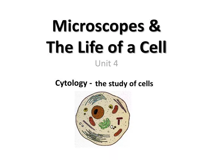
Microscopes &The Life of a CellUnit 4 the study of cells Cytology -
Microscopes Tools that helps us see things closer and with more detail
Magnification Increase in an apparent objects size
Light Microscope (LM) • 2-D images • Uses multiple lenses to increase magnification of an object • Visible light passes through the specimen and magnification lenses • Very best LM have a magnification of 2,000x • Used to observe living organisms and cells
Light Microscope Parts and Functions Holds top lens (10x magnification) Provides distance between two lenses Holds lower lenses and turns to change objective lens Supports body tube There are THREE lenses: Scanner—4x magnification Low Power—10x magnification High Power—40x magnification Supports slide Holds slide in pace Moves stage up and down to bring object into focus Controls amount of light entering slide Moves stage up and down SLIGHTLY to bring object into focus Provides light to shine on slide Supports microscope
Stereomicroscope (Dissecting Microscope) • 3-D image using light and lenses • Commonly used to study the surface of solid specimens • Lowest magnification of all microscopes
Electron Microscope (EM) • Uses a beam of electrons to create image • Can magnify up to 2 million times
Transmission Electron Microscope (TEM) • Beam of electrons passes through an ultra thin piece of specimen
Scanning Electron Microscope (SEM) • Beam of electrons scans the surface of specimen to create topographical picture of specimen How an SEM works!
Microscope Drawings • Always use pencil to draw! TITLE Amoeba cell membrane nucleus 100x TOTAL MAGNIFICATION = eyepiece x objective lens
The Cell The basic unit of life
4 mm cube Cell Diversity Cells are complex and vary in size, shape, and function (their job) Size • As a cell grows, its volume increases more rapidly than its surface area. • Surface area is too small for cell to receive materials fast enough to meet the needs of the cell • As a result, the cell divides Area = 96 mm2 Vol = 64 mm3 Ratio 1.5 / 1 Area = 384 mm2 Vol = 64 mm3 Ratio 6/1
Shape • Relates to a cells function (job) • Examples: • A flat shape of dead skin cells is well suited for covering the body surface • The long, thin, threadlike shape of nerve cells is well suited for transmitting messages through the body
Development of the Cell Theory • Robert Hooke (Great Britain)- 1665 • Examined cork (bark of oak tree) • Discovered and named “cells” • Reminded him of the small rooms in dormitories and prisons • Actually was seeing dead cell wall remains
Anton Van Leeuwenhoek (Holland- 1673) • First to describe living cells – microorganisms • Schleiden (Germany – 1838) • Determined that all PLANTS were made out of cells • Schwann (Germany – 1839) • Determined that all ANIMALS were made out of cells • Virchow (Germany – 1859) • Observed and described mitosis (cell reproduction) • Determined that cells arise from other cells
The Cell Theory • All living things are composed of one or more cells. • Cells are the basic unit of structure and function in all organisms. • All cells come from preexisting cells through cell division.
Organisms are divided into TWO groups based on cell type: • Prokaryotic cells • Eukaryotic cells
PROKARYOTIC CELLS • Lack a nucleus and membrane-bound organelles. • Unicellular. • Fossils date to 3.5 billion years ago. • Small, simple cells. • All prokaryotes are bacteria.
EUKARYOTIC CELLS • Have a nucleus and membrane-bound organelles. • Some are unicellular, most are multicellular. • Fossils date to 1.7 billion years ago. • Large, highly organized cells. • Plants, animals, protists and fungi are eukaryotes.
Why do cells have organelles? Division of labor into separate compartments (organelles) makes cell activities more organized and efficient. This is made possible by the presence of lipid membranes that form boundaries around each organelle (membrane-bound organelles).
Nucleus • Control center because it contains DNA • Surrounded by the nuclear membrane • Nuclear membrane has pores to allow things to enter/leave nucleus nucleus
CHROMOSOMES DNA that is tightly wound and organized Present during cell division only DNA Exists in Two Different Forms CHROMATIN • Loose, uncoiled strands of DNA that can be read by the cell • Present inside the nucleus during the normal, everyday life of the cell
Plasma (Cell) Membrane • Boundary of cell composed of mostly lipids; contains some protein channels for transport • Maintains homeostasis of the cell by controlling the movement of materials in and out outside of cell inside of cell
Cell Wall • Rigid outermost layer of plant cells surrounding the cell membrane • Composed of long chains of cellulose • Gives plant cells strength and support. • NOT present in animal cells
Cytoplasm • Jelly-like material that fills the cell, completely surrounding organelles cytoplasm
Chloroplast • Captures sunlight energy and uses it to make food in the form of glucose (PHOTOSYNTHESIS) • Present in LEAF cells • Contain chlorophyll pigment chloroplasts
Plastids Plant organelles that store food or pigment molecules. leucoplasts in potato cells (stained) chromoplasts in red pepper cells
Mitochondria Break down food molecules (glucose) to release usable ENERGY for all cell activities. Usable energy in cells is called ATP.
Vacuole • Fluid-filled area used for storage of water, or solutions containing food, enzymes, or waste • Plant cells have one large vacuole; animal cells have numerous small vacuoles
Ribosomes • Small, round organelles that produce proteins using genetic information from the nucleus • Can be scattered throughout the cytoplasm or bound to the Rough ER ribosomes
Nucleolus • Circular structure inside the nucleus • Responsible for making ribosomes Nucleolus Nucleus
Endoplasmic Reticulum (ER) • System of tubes and channels • Rough ER has ribosomes for protein production • Smooth ER produces lipids; also breaks down toxic substances rough ER smooth ER
Golgi Apparatus • Packaging factory for newly made lipids, proteins, and other molecules • Ships materials in vesicles (small, circular packages) to their final destination vesicles
Lysosome Contains digestive enzymes to breakdown: a) food b) old cell parts c) waste d) invading viruses and bacteria e) the cell itself lysosomes
Cytoskeleton • Network of protein fibers that gives the cell structure and support • Anchors the organelles in the cytoplasm • May assist in the locomotion of the entire cell and/or movement of organelles within the cytoplasm cytoskeleton
Centrioles • Pair of cylindrical structures that assist in cell reproduction (division) • Present in animal cells only
Flagella are longer than cilia and less numerous. Their whip-like motion moves a cell from place to place. Cilia and Flagella • Cilia are numerous short hair-like extensions that exist on the surface of some cells. • They often help move substances across a cell’s surface.