Download
1 / 48
771 likes | 1.38k Views
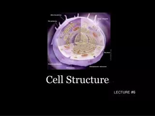
Cell Structure. Cell Theory. All organisms are made of cells The cell is the simplest collection of matter that can live Cell structure is correlated to cellular function All cells are related by their descent from earlier cells. Organisms and Cells.

E N D
Cell Theory • All organisms are made of cells • The cell is the simplest collection of matter that can live • Cell structure is correlated to cellular function • All cells are related by their descent from earlier cells
In 1665, the English scientist Robert Hooke coined the term “cellulae” for the small box-like structures he saw while examining a thin slice of cork under a microscope.
Cells were discovered in 1665 by Robert Hooke. Early studies of cells were conducted by - Mathias Schleiden (1838) - Theodor Schwann (1839) Cell Theory
Cell Theory • A unifying concept in biology • Originated from the work of biologists Schleiden and Schwann in 1838-9 • States that: • All organisms are composed of cells • German botanist Matthais Schleiden in 1838 • German zoologist Theodor Schwann in 1839 • All cells come only from preexisting cells • German physician Rudolph Virchow in 1850’s • Smallest unit of life
Cell size is limited. -As cell size increases, it takes longer for material to diffuse from the cell membrane to the interior of the cell. Surface area-to-volume ratio: as a cell increases in size, the volume increases 10x faster than the surface area
Cell Size • Most much smaller than one millimeter (mm) • Some as small as one micrometer (mm) • Size restricted by Surface/Volume (S/V) ratio • Surface is membrane, across which cell acquires nutrients and expels wastes • Volume is living cytoplasm, which demands nutrients and produces wastes • As cell grows, volume increases faster than surface • Cells specialized in absorption modified to greatly increase surface area per unit volume
Sizes of Living Things Most cells are microscopic 5-100 microns Length of nerve cell 1 meter (m) Largest single cell Ostrich egg
Microscopes are required to visualize cells. Light microscopes can resolve structures that are 200nm apart. Electron microscopescan resolve structures that are 0.2nm apart.
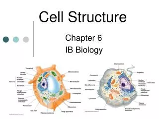
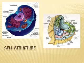
All cells have certain structures in common. 1. genetic material – in a nucleoid or nucleus 2. cytoplasm – a semifluid matrix 3. plasma membrane – a phospholipid bilayer
Prokaryotic Cells • Lack a membrane-bound nucleus • Structurally simple • Two domains: • Bacteria • Three Shapes • Bacillus (rod) • Coccus (spherical) • Spirilla (spiral) • Archaea • Live in extreme habitats
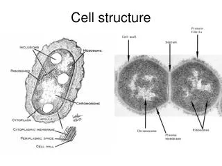
Prokaryotic Cells Prokaryotic cells possess -genetic material in the nucleoid -cytoplasm -plasma membrane -cell wall -ribosomes -no membrane-bound organelles
Prokaryotic Cells Prokaryotic cell walls -protect the cell and maintain cell shape Bacterial cell walls -may be composed of peptidoglycan -may be Gram positive or Gram negative Archaean cell walls lack peptidoglycan.
Prokaryotic Cells Flagella -present in some prokaryotic cells -used for locomotion -rotary motion propels the cell
Eukaryotic Cells -possess a membrane-bound nucleus -are more complex than prokaryotic cells -compartmentalize many cellular functions within organelles and the endomembrane system -possess a cytoskeleton for support and to maintain cellular structure
Nucleus -stores the genetic material of the cell in the form of multiple, linear chromosomes -surrounded by a nuclear envelope composed of 2 phospholipid bilayers -in chromosomes – DNA is organized with proteins to form chromatin
Ribosomes -the site of protein synthesis in the cell -composed of ribosomal RNA and proteins -found within the cytosol of the cytoplasm and attached to internal membranes
Endomembrane System • Restrict enzymatic reactions to specific compartments within cell • Consists of: • Nuclear envelope • Membranes of endoplasmic reticulum • Golgi apparatus • Vesicles • Several types • Transport materials between organelles of system
Rough endoplasmic reticulum (RER) -membranes that create a network of channels throughout the cytoplasm -attachment of ribosomes to the membrane gives a rough appearance -synthesis of proteins to be secreted, sent to lysosomes or plasma membrane
Smooth endoplasmic reticulum (SER) -relatively few ribosomes attached -functions: -synthesis of membrane lipids -calcium storage -detoxification of foreign substances
Golgi apparatus -flattened stacks of interconnected membranes -packaging and distribution of materials to different parts of the cell -synthesis of cell wall components
Lysosomes -membrane bound vesicles containing digestive enzymes to break down macromolecules -destroy cells or foreign matter that the cell has engulfed by phagocytosis
Vacuoles -membrane-bound structures with various functions depending on the cell type -There are different types of vacuoles: -central vacuole in plant cells -contractile vacuole of some protists -vacuoles for storage
Vacuoles • Membranous sacs that are larger than vesicles • Store materials that occur in excess • Others very specialized (contractile vacuole) • Plants cells typically have a central vacuole • Up to 90% volume of some cells • Functions in: • Storage of water, nutrients, pigments, and waste products • Development of turgor pressure • Some functions performed by lysosomes in other eukaryotes
Microbodies -membrane bound vesicles -contain enzymes -not part of the endomembrane system -glyoxysomes in plants contain enzymes for converting fats to carbohydrates -peroxisomes contain oxidative enzymes and catalase
Peroxisomes • Similar to lysosomes • Membrane-bounded vesicles • Enclose enzymes • However • Enzymes synthesized by free ribosomes in cytoplasm (instead of ER) • Active in lipid metabolism • Catalyze reactions that produce hydrogen peroxide H2O2 • Toxic • Broken down to water & O2 by catalase
Mitochondria -organelles present in all types of eukaryotic cells -contain oxidative metabolism enzymes for transferring the energy within macromolecules to ATP -surrounded by 2 membranes -smooth outer membrane -folded inner membrane with layers called cristae
-matrix is within the inner membrane -intermembrane space is located between the two membranes -contain their own DNA
Chloroplasts -organelles present in cells of plants and some other eukaryotes -contain chlorophyll for photosynthesis -surrounded by 2 membranes -thylakoids are membranous sacs within the inner membrane -grana are stacks of thylakoids
Cytoskeleton -network of protein fibers found in all eukaryotic cells -supports the shape of the cell -keeps organelles in fixed locations -helps move materials within the cell Cytoskeleton fibers include -actin filaments – responsible for cellular contractions, crawling, “pinching” -microtubules – provide organization to the cell and move materials within the cell -intermediate filaments – provide structural stability
The Cytoskeleton: Microtubules • Hollow cylinders made of two globular proteins called a and b tubulin • Spontaneous pairing of a and b tubulin molecules form structures called dimers • Dimers then arrange themselves into tubular spirals of 13 dimers around • Assembly: • Under control of Microtubule Organizing Center (MTOC) • Most important MTOC is centrosome • Interacts with proteins kinesin and dynein to cause movement of organelles
Cell Movement Cell movement takes different forms. -Crawling is accomplished via actin filaments and the protein myosin. -Flagella undulate to move a cell. -Ciliacan be arranged in rows on the surface of a eukaryotic cell to propel a cell forward. The cilia and flagella of eukaryotic cells have a similar structure: -9+2 structure: 9 pairs of microtubules surrounded by a 2 central microtubules -Cilia are usually more numerous than flagella on a cell.
Extracellular Structures Extracellular structures include: -cell walls of plants, fungi, some protists -extracellular matrix surrounding animal cells
Cell Wall -present surrounding the cells of plants, fungi, and some protists -the carbohydrates present in the cell wall vary depending on the cell type: -plant and protist cell walls - cellulose -fungal cell walls - chitin
Extracellular matrix (ECM) -surrounds animal cells -composed of glycoproteins and fibrous proteins such as collagen -may be connected to the cytoplasm via integrin proteins present in the plasma membrane
Electron Microscope • Resolves fine structure of the cell • Uses an electron beam rather than a light beam • Relationship between limit of resolution and wavelength applies for any form of radiation • Resolving power of electron microscope is 0.1 nm • Source of illumination is a filament (cathode) that emits electrons at the top of the column • Since electrons are scattered by collisions with air molecules, column must be under a vacuum
How is contrast achieved in the electron microscope? • Specimen is stained with an electron dense material • Some of the electrons passing through the specimen are scattered by structures stained with electron dense material • Others pass through parts of the cell not stained to form an image on a phosphorescent screen • Because the scattered electrons are lost from the beam, the stained regions show up as dark • Thus the image is a montage of light (non stained) and dark (stained) regions
Preparation of Specimens • Preserved by fixation • 1st, glutaraldehyde • Covalently cross-links proteins • 2nd, osmium tetroxide • Binds to and stabilizes lipid bilayers and proteins • Tissue dehydrated, permeated with a polymerizing resin & sectioned into ultra-thin sections • 50 – 100 nm thick (1/200 thickness of a cell)
Sections stained with electron-dense material (e.g uranyl acetate) to achieve contrast • How does this work? • Tissue composed of atoms of low atomic number (e.g. carbon, oxygen, nitrogen, hydrogen) • To make them visible impregnated with salts of heavy metals.
Immunogold ElectronMicroscopy • Used to visualize specific proteins • Incubate thin section with primary antibody • Then incubate with secondary antibody to which colloidal gold has been attached • Gold is electron dense and shows up as black dots
Electron Microscopyof Metal-Shadowed Samples • The transmission electron microscope (TEM) can be used to resolve individual macromolecules on the surface of the specimen • Thin film of heavy metal (e.g. platinum) is evaporated onto the dried specimen
Electron MicroscopyofMetal-Shadowed Samples • For thick samples (e.g. cells), the organic material is dissolved away after shadowing • Only the thin metal replica of the surface is left • This is thin enough for the electron beam to penetrate.
Metal-shadowed electron micrograph of a freeze-fractured membrane