Download
1 / 71
720 likes | 833 Views
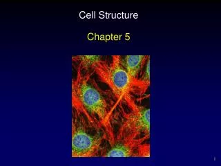
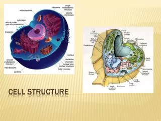
Cell structure. Fields of study Cytology Histology Pathology. Levels of Organization Cells Tissues Organs Organ systems Individual. Cells vary in size and shape according to location and function.

E N D
Fields of study Cytology Histology Pathology Levels of Organization Cells Tissues Organs Organ systems Individual
Cells vary in size and shape according to location and function. Cells vary in internal structure depending upon their function. Cells vary in their life history, e.g., rates of cell renewal. Neuron Lining cell Macrophage Muscle cell Neuron Enterocyte
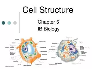
Nucleus Nuclear envelope Cytoplasm Organelles Plasma membrane Plasma membrane Major compartments of the cell Cytoplasm - provides an aqueous matrix containing the internal structures of the cell and the cytosolic metabolic pathways, e.g., glycolysis Nucleus – houses the DNA, production of mRNA, initial ribosome assembly
Cell membranes • General concepts • a. Intracellular – forms organelles • b. Cell surface – plasma membrane • c. All membranes have a similar structure, • referred to as unit membranes • d. Appears tri-laminar by electron microscopy • due to the deposition of osmium Unit membrane • Fluid mosaic model of membrane structure • a. Two phospholipid layers (phospholipid bilayer) • b. Associated proteins • i. Integral • ii. Peripheral
I I I I P P Plasma membrane Integral membrane proteins Insert into one leaflet Transmembrane (channels, receptors) – Span both leaflets Peripheral membrane proteins
Freeze fracture Plasma membrane
Freeze fracture Exposed membrane surface is “replicated” by coating with a thin layer of metal. Replica is view with a transmission EM
EM 150,000x Plasma membrane LM 1,000x EM 10,000x
Glycocalyx (PAS positive)
Functions of the plasma membrane • Transport • Diffusion - depends on the hydrophobicity, size, charge, presence of channel proteins. Passive diffusion Facilitated diffusion – uses transmembrane proteins • Active transport (energy dependent, e.g., ATP) • Vesicular transport (macromolecules/cell debris/microbes)
Vesicular Transport Endocytosis - internalization of small membrane vesicles from the plasma membrane; involves vesicles < 1.0 micron a) Pinocytosis (“cell drinking”) - uptake of fluid by a continuous process; membrane recycling. b) Receptor-mediated endocytosis – requires receptor-ligand binding for vesicle formation and internalization.
Phagocytosis Pinocytosis vs phagocytosis Pinocytosis 100,000x Phagocytosis (“cell eating”) Ingestion of large particles,e.g., bacteria; restricted to macrophages and leucocytes; involves particles > 1.0 micron
Receptor-mediated endocytosis Triskelion LDL and the LDL receptor Transferrin Clathrin Clathrin-coated vesicle
Vesicular Transport Exocytosis - Fusion of cytoplasmic vesicles with the plasma membrane and release of the vesicle contents to the outside of the cell a) Constitutive exocytosis - continuous process, e.g., renewal of plasma membrane, continuous secretion b) Regulated exocytosis - requires extracellular signal for vesicle fusion and release, e.g., hormone secretion and neurotransmitter release (“secretion on demand”)
Transcytosis Transcytosis - Uptake of material on one side of a cell followed by transport and release from the opposite surface. Vessel lumen Endothelial cell Connective tissue Common in endothelial cells which line blood vessels. Also a mechanism used by some cells for protein trafficking to apical or basal membrane domains.
Cell junctions Cell contacts established by integral membrane proteins Best developed in epithelial tissues which are present at body surfaces 1 1. Seal adjacent Tight junctions cell membranes establish apical and basal domains 2. Attach and anchor cells Adherent junctions 3. Provide channels for Gap junctions ionic continuity between cells 2 2 2 3 On the CD these structures are included with Epithelium
Tight junction (zonula occludens) Belt-like barrier around the apex of the cell Close apposition of membrane proteins, exclusion of intercellular space
Zonula occludens - Excludes luminal contents from intercellular space - Prevents migration of proteins within the cell membrane, maintains apical and basolateral domains - Maintains cell polarity
Adherent junctions - Anchor cells to each other or to the basal lamina • No membrane fusion • Types of adherent junctions • Spot desmosome (macula adherens) • Hemidesmosome • Belt desmosome (zonula adherens)
Desmosome (Macula adherens) Adherent junctions
ZO ZA D Junctional complex • Components • Zonula occludens (ZO) • Zonula adherens (ZA) • Desmosome (D) • Epithelial tissues only
Gap junctions Composed of six transmembrane proteins clustered like a rosette called a connexon. Adjacent connexons form a pore between cells Provide metabolic and electrical continuity between cells (coupling)
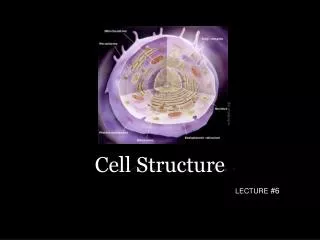
Cell compartments Nucleus Cytoplasm Endoplasmic reticulum Golgi apparatus Lysosomes Mitochondria Peroxisomes
Nucleus Eukaryotic cells Provides sequestered compartment for DNA Produces pre-ribosomal particles Site of messenger RNA synthesis
Nucleus Electron microscopy 5,000x Light microscopy (H & E) 1,000x
Nuclear envelope
EM 18,000x (Freeze fracture) Nuclear envelope and nuclear pores EM 65,000x
Electron micrograph Nuclear pores Regulate passage of materials in and out of the nucleus Corbett & Krebber, 2004 Nuclear pore proteins are common targets in autoimmune diseases, e.g., biliary cirrhosis and inflammatory bowel disease
Nucleolus Ribosomal RNA synthesis Ribosomal subunit assembly Not surrounded by a membrane LM 1000x EM 12,000x
Regions of the nucleolus 1. Nucleolar organizing centers (fibrillar centers) Site of DNA encoding rRNA 2. Pars fibrosa (dense fibrillary component) Contains rRNA transcripts 3. Pars granulosa Site of ribosomal particle assembly EM 40,000x
ChromatinDNA + Protein (mostly histones) Euchromatin Transcriptionally active Dispersed Pale staining Heterochromatin Transcriptionally inactive Condensed Dark staining LM 1,000x
Light microscopy (H & E, 1,000x) Electron microscopy (6,000x) Most nuclei show a mixture of heterochromatin and euchromatin
Endoplasmic reticulum Rough endoplasmic reticulum (RER) Smooth endoplasmic reticulum (SER)
Rough endoplasmic reticulum Composed of flattened membranous sacs (unit membranes) Possesses ribosomes on the outer cytoplasmic surface Continuous with the nuclear envelope Site of protein synthesis and some phospholipid synthesis
Rough endoplasmic reticulum Light microscopy (H&E, 1,000x) Electron microscopy 15,000x
Smooth endoplasmic reticulum Tubular membranes continuous with RER No ribosomes Functions: - Site of triglyceride, cholesterol and steroid hormone synthesis - Detoxification - Calcium storage
Smooth endoplasmic reticulum 30,000x 10,000x
Ribosomes Two subunits, composed of rRNA and protein Site of protein synthesis (translation) Distribution - Free in the cytoplasm (polysomes) - Attached to the endoplasmic reticulum (RER) Polysomes RER EM 75,000x; 250,000x EM 20,000x
Sites of protein synthesis Polysomes Cytoplasmic proteins, e.g., glycolytic enzymes and cytoskeletal proteins Mitochondrial proteins Peroxisomal proteins Nuclear proteins, e.g., transcription factors ribosomal proteins and histones Rough endoplasmic reticulum Proteins for secretion Lysosomal enzymes Integral membrane proteins Resident proteins for ER and Golgi
Sites of protein synthesis Polysomes RER
Golgi apparatus Flattened membranous sacs Usually located near the nucleus No direct continuity with ER Site of post-translational processing and vesicle packaging Receives products synthesized in the ER
Golgi apparatus Cis face (forming face) Trans Golgi network Mid-Golgi region Trans face (maturing face)
Golgi apparatus
Golgi apparatus – Classical model Transition/transfer vesicles Forming face/cis face Maturing face/trans face Trans-Golgi network EM 75,000x
Golgi apparatus – Classical model Transition/transfer vesicles Forming face/cis face Maturing face/trans face Trans-Golgi network EM 75,000x
Golgi apparatus – Classical model Transition/transfer vesicles Forming face/cis face Maturing face/trans face Trans-Golgi network EM 75,000x
Golgi apparatus – Classical model Transition/transfer vesicles Forming face/cis face Maturing face/trans face Trans-Golgi network EM 75,000x
Cis-golgi network Trans-golgi network Cisternal progression model