Download
1 / 63
690 likes | 1.63k Views
1 . This cow has ingested ethylene glycol anti-freeze and became a downer, just like in milk fever. What is the cause of acidosis in this case? A. ethylene gas B . alcohol dehydrogenase C . glycolic acid D . acetic acid. 2 . Histopath sections showed oxalate

E N D
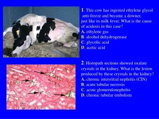
1. This cow has ingested ethylene glycol anti-freeze and became a downer, just like in milk fever. What is the cause of acidosis in this case? A. ethylene gas B. alcohol dehydrogenase C. glycolic acid D. acetic acid 2. Histopath sections showed oxalate crystals in the kidney. What is the lesion produced by these crystals in the kidney? A. chronic interstitial nephritis (CIN) B. acute tubular necrosis C. acute glomerulonephritis D. chronic tubular embolism
3. This kidney belonged to a cat that was showing signs of polyuria and later on progressed to anuria indicating severe nephron damage. Previously the cat was diagnosed to be positive to Feline Infectious Peritonitis. There is still hope that kidney function can be restored if the following lesions are not observed: A. glomerulotubular necrosis and interstitial nephrosis B. interstitial hemorrhage and tubular edema C. damaged basement membrane and fibrosis D. infarct and embolism
4. If this is the kidney and blood of a cow that ingested too much bracken fern (Pterygium aquilinum), a lot of damage will happen in the kidney due to the plant alkaloid toxin quercetin. The diagnosis for this disease is: A. toxic glomerulonephritis C. bovine enzootic hematuria B. hemoglobinuric nephritis D. grass tetany 5. If the cow will not immediately die and will continue to eat the bracken fern, further pathological lesions will occur in the urinary bladder, which is histopathologically described as: A. urinary bladder myoma B. squamous cell carcinoma C. hemorrhagic lymphoma D. urinary osteochondroma
6. The cat from which this kidney was taken obviously ingested a nephrotoxic agent. Select the item NOT TRUE for this case: A. low creatinine B. high BUN 7. Urinalysis confirmed the presence of cast. This cast formation is indicative of: A. severe tubular necrosis B. nephrotic syndrome C. toxic nephritis D. renal sarcoma
8. This horse had laminitis and was treated with phenylbutazone. Instead of getting better the horse died and the kidney showed papillary necrosis secondary to ischemia. Select the statement NOT TRUE for this condition: A. The phenylbutazone interfered with production of renal prostaglandins. B. The alteration in prostaglandins caused vasoconstriction and loss of perfusion to the distal tissues of the kidney. C. The phenylbutazone produced toxic glomerulonephritis. D. The vasocontriction caused the papilla to become ischemic and then necrotic.
9. The dog had necrotic brown tissue in the kidney (white arrows). A sample of urolith is shown in the right picture. Select the statement that is NOT TRUE in this case: A. The necrotic tissue can cause ureteral obstruction. B. Pieces of necrotic tissue can initiate urolith formation. C. Necrotic tissue is the cause of nephroblastoma. D. A nidus is necessary for a calculus to form.
10.Renal failure is a general term that results from lesions causing total kidney damage and loss of function leading to imbalance Ca:P ratio and uremia. Name the specific lesions associated with renal failure by filling in the blanks for the following pictures: A. fibrous _____________ B. endocardial __________ C. fibrinous ___________ F. ulcerative glossitis & ____________necrosis E. ulcerative _________ D. gastric ________ & hemorrhage
11. This dog shows signs of typical NEPHROTIC SYNDROME. Enumerate the 4 components of NEPHROTIC SYNDROME: A. _________________ B. _________________ C. _________________ D__________________
12. It is contraindicated to give i.v. fluid to animals with nephrotic syndrome because of this danger. What bad pathological effect will occur if i.v. fluid is given to animals with nephrotic syndrome? __________________________ 13. Another consequence of nephrotic syndrome is thrombosis. Can you explain the pathogenesis of thrombosis in animals with nephrotic syndrome? ___________________________ ___________________________ ___________________________
14.This is a female Dalmatian with urate calculi as shown in the lower picture. Select the statement NOT TRUE for Dalmatians: A.They excrete large quantities of uric acid due to a deficiency of uptake of uric acid by the liver. B.Urate calculi formation affects only female Dalmatians. C.They secrete uric acid which is less soluble than allantoin D. There is hyperconcentration of uric acid in the urine and formation of urate calculi.
15.These are 2 cases in cats showing hemorrhagic urinary bladder due to calculus obstruction of the urinary tract as a cause of Feline Urologic Syndrome (FUS). This FUS is most commonly observed in what sex of the cat ? A. female B. male
16.The diagnosis for this dog’s urinary bladder is emphysematous cystitis (Fanconi syndrome). What is the cause of this inflammation? A. Canine distemper B. Leptospirosis C. Diabetes mellitus D. Rat poison (dicoumarol)
17.This is a polycystic kidney from a dog. What is the best description for the gross appearance of this kidney? A. moth-eaten appearance B. cavernous appearance C. sponge-like appearance D. swiss cheese-like appearance
18.This is the kidney from a German Shepherd dog with renal cystadenocarcinoma. What is the pathogenesis of this tumor? A. acquired - defect in the embryogenesis B. hereditary - loss of tumor suppressor gene C. infectious - induced by Canine Distemper virus D. toxic - induced by heavy metal (Lead) poisoning
19.This is the kidney from a frog with renal carcinoma. T denotes tumor areas and N are normal tubules.What is the etiology of this tumor in frogs? A. acquired - defect in the organogenesis B. hereditary - duplication of dominant oncogene C. infectious - induced by herpes virus D. toxic - induced by heavy metal (Mercury) poisoning
20. This is the kidney of a cat with renal lymphosarcoma. What disease in the cat should you consider as a rule-out for the differential diagnosis of this tumor? ___________________________________________
21. This is the urinary bladder from a sheep. Name the 3 most possible tumors that can cause this condition: 1. _____________________________ 2. _____________________________ 3. _____________________________
22.This disease in sheep (and also in goats) is caused by contagious ecthyma virus. The following are similar descriptive terms for this disease EXCEPT ONE: A. exudative cheilitis B. orf C. nodular gingivitis D. proliferative stomatitis
23.These are classical pictures of “lumpy jaw” in cattle every veterinary student should be familiar with. A. What is the causative agent ?__________________ B. What is the morphological diagnosis ?___________ ______________________________________
24.This is a photo of a cow that swallowed something too big and it got stuck in the esophagus. The cow died. There are 4 points along the esophagus that are narrowest and prone to choke. Select the point that is NOT PRONE to choke: A. over the larynx B. thoracic inlet C. at the midpoint D. over the heart base E. just in front of the diaphragmatic inlet
25. TRUE or FALSE. In cases of LIPIDOSIS, the distinct margins on the vacuoles are a giveaway and this is a criteria for differentiation from vacuolar change due to glycogen, which do not have crisp borders as lipid vacuoles do.
26. This dog is "headpressing" due to hepatic encephalopathy. On necropsy, the dog had severe hepatic cirrhosis or "end-stage" liver disease (right picture) TRUE or FALSE: This condition is due to glucoronic acid retention and retention of toxic oxalates normally removed from the portal blood by the liver.
27. An area of the liver is shown with classical histopath lesion of hepatic necrosis. Select the specific nuclear change in this picture: A. pyknosis B. karyorrhexis C. karyolysis
28. This cocklebur(Xanthium strumarium) if accidentally ingested by animals can cause hepatic necrosis. What kind of hepatic necrosis is produced by this plant? A. centrilobular necrosis B. mid-zonal necrosis C. periportal necrosis
29.A section of the liver with bile duct hyperplasia. TRUE or FALSE: The first 2 reactions to liver injury are REGENERATION and FIBROSIS. The 3rd reaction is BILE DUCT HYPERPLASIA.
Low power magnification High power magnification 30.A stray puppy caged in the animal shelter showed jaundice, swollen and reddened tonsils. Palpation showed a greatly swollen liver. It died and necropsy confirmed the swollen liver. The histopath is shown above. Since it was a stray puppy it had no vaccination. What is your diagnosis ?______________________________
A B 31. Livers A and B are from an adult cattle. Abscess is caused by by a specific bacteria in liver A and a secondary infection caused the lesion in liver B. Can you name the primary and secondary bacterial infection in this 2 livers? A. Liver A caused by ___________________________ B. Liver B caused by ___________________________
A B 32. A foal 3 weeks old died. Necropsy showed focal hepatitis and necrosis (A) and high magnification showed bacteria (B) arranged like Chinese character. A. What is the name of the disease ?_________________ B. What is the bacterial etiology ?___________________
33. A litter of pigs showed unthriftyness. You necropsied one piglet and you saw lots of Ascaris suum in the small intestine. Multifocal lesions were found in the liver as shown above. Lung was apparently normal. A. What is the layman’s term for the multifocal lesion in the liver ? ____________________ B. What is the cause of these multifocal liver lesion ?______________________
34. This is a Chinese Sharpei breed of dog. It is susceptible to this kind of liver disorder. Histopath of liver stained with Congo red under polarized light showed birefringence (right picture): A. liver carcinoma B. bile duct hyperplasia C. porto-caval shunt D. liver amyloidosis
35. A large number of rabbits were affected acutely and died suddenly. The characteristic signs of blood oozing out from the nostril was observed (epistaxis). A. What is the diagnosis ?_______________________ B. What is the etiology? ________________________
36.This disease in cattle particularly affects the liver, as shown in the left picture. The histopath on the right shows the typical granulomatous inflammation surrounded by giant cells. A. What is your diagnosis ?_______________________ B. What is the etiology ? _________________________ C. What special stain can confirm presence of the etiological agent in the histopath section ? ___________________
37. Moldy corn was accidentally fed to swine and the animals died due to aflatoxicosis affecting the liver, as shown in the right picture. What kind of necrosis is produced by aflatoxin in the liver ?: A. midzonal necrosis B. peripheral necrosis C. centrilobular necrosis
38.This picture shows pancreas with neoplastic growths. TRUE or FALSE: If the neoplasm arises from the endocrine cells, specifically the beta cells of the islets, it is termed an insulinoma, if benign; or islet cell carcinoma, if malignant.
39. A dog was suffering from severe abdominal pain and died. Necropsy showed that the pain originated from the diseased pancreas shown in the picture above. Can you pick out the 2 classical lesions from the list below ? A. pancreatic abscesses and hemorrhages B. pancreatic fat necrosis and saponification C. pancreatic amyloidosis and gangrene D. pancreatic hyperplasia and adenoma
B A C 40.This pig (A) was suffering from severe diarrhea and died because of dehydration. Histopath showed villous atrophy (B) of the small intestine which was confirmed by immunohistochemistry for the causative agent (C). Name the 2 most probable diagnosis ? _________
41.The two lesions depicted in this dog are plaque and gingivitis. How do these lesions result in gingival hyperplasia? A. Gingivitis and plaque result in several types of neoplasia and gingival hyperplasia is an intermediate stage. B. The gingivitis and plaque are caused by bacteria that cause edema and gingival hyperplasia. C. Gingivitis and plaque lead to periodontal disease and causes proliferation of fibroblasts in the gingiva. D. Gingivitis and plaque stimulate macrophages to proliferate and these make the gums swell.
42.This is a classical lesion in the esophagus of a calf. Can you identify the lesion ? HINT: the esophagus is lined by stratified squamous epithelium. The lesion is: ______________________________
43. It was a hot summer day when you were called by a livestock farmer because he found his cattle dead on the field with bloated abdomen. The origin of gas accumulation in ruminants that die must be identified as either antemortem or postmortem. However, based from your experience as seen in the picture on the right, you are sure the cause of the bloat is antemortem, not postmortem. A. Why was the bloat antemortem ? _______________________ B. Explain the pathogenesis of this bloat ___________________ ____________________________________________________
B A 44. The puppy had bloody diarrhea with characteristic fish odor. Treatment was attempted but the poor puppy died. Necropsy showed the small intestine with reddened areas in segmented pattern (A). The serosal surface had “ground glass appearance” due to fibrin exudation.Histopath exam confirmed your suspicion when you saw the distinct round blue-to-red bodies within a vacuolated nucleus that has marginated chromatin (B). A. What is the diagnosis ? _________________________________ B. Explain the pathogenesis of leukopenia in infected dogs as a complicating factor of this disease. _______________________________________ _________________________________________________________
C A D B 45. The piglets were dehydrated due to watery diarrhea (A). Necropsy showed dilated and fluid- filled instestine (B). Histopath showed villous atrophy (C). Electron microscopy identified the causative agent with characteristic morphological appearance (D). WHAT IS THE DIAGNOSIS ? _______________________
B A 46. The causative agent gets into enterocytes, mostly crypt cells, of the ileum and causes them to proliferate. Affected gut (A) is on the top; normal is below There is a definite "cerebral" pattern to the intestinal wall. Histopath (B) shows the lymphoid tissues of Peyer’s patches underneath the thick polypoid proliferating mucosa, not in an orderly form of epithelium. This form of PPE (PorcineProliferative Enteropathy) is known as porcine intestinal adenomatosis (PIA) What is the ETIOLOGY ?__________________________________
A B 47. Grossly, the disease can be hemorrhagic (A), or more fibrinous (B) Either way, the pig has diarrhea and is dehydrating out its hind end. The feces are reported to be quite characteristic because of all the eclectic contents. They are described as being gray and greasy, and often flecked with fibrin or blood. The pigs, because of the chronic diarrhea, are often described as being "slab-sided" because they lose the nice back fat that makes them appear rounded and slightlyroly-poly. A. What is the diagnosis ? _______________________ B. What is the etiology ? ________________________
C A B 48.A dairy farmer called you because some of his cows were suffering from diarrhea. One animal is shown in picture A with unthrifty appearance and with swollen cervical lymph nodes. Picture B shows affected and normal intestine above and below, respectively. Take note of the thick corrugated intestinal mucosa. Picture C shows the big ballooned macrophages in the intestinal mucosa and picture D the same section stained with acid-fast showed that the big ballooned macrophages contained acid-fast bacilli. D A. What is the name of the disease ? B. What is the etiology of this disease ?
A B C 49.This is usually a systemic disease in dogs and other animals, with the worst lesions in the lung. Sometimes there are lesions in the intestines as well (A). The lamina propria gets chock-full of macrophages filled with the organism and there is backup of lymph flow (picture A, arrows). On cut section, the lamina propria is markedly expanded (B). There is a macrophage in the center, filled with the organism (C). WHAT IS THE ETIOLOGY ? ____________________________
C A B 50. A very thin puppy was brought to your clinic with very pale mucous membrane. Fecal examination showed the presence of lots of parasite eggs (A). You tried to save the poor puppy but it finally died. Necropsy examination showed bloody small intestine (B) and closer examination revealed adult worms in large numbers (C). What is the diagnosis for this case ? _____________________
51. - 52. You are called upon by the owner of a 1000 sow level farm to investigate a disease outbreak. These are your preliminary findings: • Pigs are found dead in the grower and finisher • Some live pigs are lame and have swollen joints • Purple, elevated skin lesions • Necropsy: vegetative endocarditis, petechial hemorrhage on kidney • Give 2 probable diagnosis for this disease ___________________________ ___________________________
53. Which sentence is NOT TRUE about Streptococcus infection in pigs? • Causes meningitis and polyserositis in piglets • Aujezky’s disease, Glasser’s disease and Septicaemic E. coli are included as differential diagnosis • It is not zoonotic • Caused by gram positive, hemolytic Streptococcus suis type 2
54. Which lesion is NOT caused by Vitamin E deficiency? • Mulberry heart disease in pigs • Hepatic necrosis • Gastric ulcer d. Encephalomalacia in chickens
55. Which sentence is TRUE about Leptospirosis? • People cannot be infected by contact of infected pig urine • Abortions, stillbirth and neonatal mortality are clinical signs of chronic Leptospira pomona infection. • Infarction of the kidney is a common lesion in chronic leptopirosis • Abortion by leptospirosis does not cause inflammation in the placenta