Download
1 / 36
370 likes | 1.22k Views
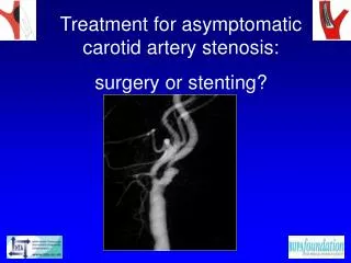
Carotid Vascular Disease: Treatment options using surgery and interventional radiology Emily Borod MS3 Carotid Vascular Disease Stroke is 3 rd leading cause of death in US (behind heart disease and cancer) Mortality from acute event is 20% 50% of patients are alive after 5 years

E N D
Carotid Vascular Disease: Treatment options using surgery and interventional radiology Emily Borod MS3
Carotid Vascular Disease • Stroke is 3rd leading cause of death in US (behind heart disease and cancer) • Mortality from acute event is 20% • 50% of patients are alive after 5 years • 4% of survivors require long-term skilled nursing care • 25% of survivors will have a second neurologic event
Signs/symptoms of carotid vascular disease • TIA (Transient Ischemic Attacks): focal neurologic defects with resolution of symptoms within 24 hours • RIND (Reversible Ischemic Neurologic Deficit): transient neurologic defects lasting 24-72 hrs • Amaurosis fugax: temporary blindness in one eye, frequently described as “curtain coming down” due to microemboli in retina • CVA (Cerebrovascular accident): neurologic deficit with permanent brain damage
Evaluating carotid disease • Duplex Doppler ultrasonography • Carotid Doppler ultrasonography • Magnetic resonance angiography (MRA) • Carotid angiography (gold standard) • Sensitivity/specificity of noninvasive tests to predict stenoses >70% is 83-86%/89-94%
Treatment options • Medical treatment (not as effective for more advanced disease) • Carotid endarterectomy (CEA) • Nonsurgical carotid revascularization using angioplasty and stenting • Treatment recommended for: • Asymptomatic pts with >60% stenosis • Symptomatic pts with >50% stenosis
Carotid endarterectomy • Performed through neck incision, usually along sternocleidomastoid muscle • Proximal and distal control of artery is obtained • While patient is heparinized, internal and external carotid arteries are clamped • Longitudinal arteriotomy is performed, carotid plaque is removed, and vessel is closed over a patch
Cardiac events Postoperative stroke Hyperperfusion syndrome Nerve injury Bleeding Infection Parotitis Re-stenosis Complications of carotid endarterectomy(perioperative mortality <0.5-3.0%, related level of expertise of surgeons)
Postoperative cardiac events • Appropriate cardiac work-up is essential • Because these patients have atherosclerotic disease in the carotids, it must be assumed that they have atherosclerotic disease elsewhere • Exercise stress testing, dobutamine echocardiography, dipyridamole imaging, or coronary catherization should be used
Postoperative stroke • Factors that contribute to postoperative stroke: • Plaque emboli • Platelet aggregates • Improper flushing • Poor cerebral protection • Relative hypotension
Hyperperfusion syndrome • Cerebral hyperperfusion is the leading cause of intracerebral hemorrhage and seizures during the first two weeks following CEA • Causes changes in low-flow carotid vascular bed • Small vessels compensate by dilating, then cannot re-constrict properly and therefore cannot protect vascular bed
Nerve injury • Nerves at risk for injury during CEA include: • Vagus nerve • Recurrent laryngeal nerve • Facial nerve • Glossopharyngeal nerve • Hypoglossal nerve • Branches of trigeminal nerve
Re-stenosis • Re-stenosis following CEA occurs in 20% of patients overall, and 2.6-10% at 5 years • Re-stenosis within 6 months is more common when smooth muscle cells are abundant in lesion and is less common when lesions are rich in lymphocytes and macrophages • Late re-stenosis results from progression of atherosclerotic disease
Predictors of mortality following CEA • Increased age • Male sex (relative risk 1.58) • Diabetes (RR 1.48) • Systemic hypertension (RR 1.31) • Smoking (RR 1.13)
Predictors of recurrence following CEA • Elevated cholesterol • Systemic hypertension (RR 1.42) • Smoking (RR 1.47)
Nonsurgical carotid revascularization • Percutaneous catheterization techniques have led to carotid angioplasty and stent placement • Less invasive (performed with local anesthesia and sedation) • Less likely to precipitate cardiac events • Technique can also be used to repair stenosis that is more cephalad
Technique used in nonsurgical carotid repair • Catheter with umbrella tip is inserted in stenotic carotid via femoral artery • Balloon is inflated to dilate artery • Stent is placed in artery to maintain patency • Filters are used to capture embolic particles
Risks of nonsurgical vascular repair • Plaques may be dislodged during procedure leading to neurologic events • Re-stenosis is common in long term follow-up (15%) and may be difficult to treat surgically • Dissection has been shown to occur in 5% of patients following stenting • More studies comparing CEA to nonsurgical repair must be completed
CEA vs stenting • Several studies have been carried out or are in progress to compare CEA and repair of carotid artery disease using interventional radiology • Because of the potentially significant and lasting damage from a stroke and the relative success of CEA, studies comparing the two treatment options have been somewhat slow to be carried out • Most of the early studies compare the two techniques in specific patient groups (i.e. elderly patients or poor surgical candidates)
WALLSTENT trial • 219 patients with symptomatic stenosis • Carotid arteries were 60-90% occluded • Patients were randomly assigned to receive CEA or angioplasty and stenting (without protective filter device) • 1-yr follow-up showed significantly higher rate of post-procedure stroke with angioplasty and stenting group compared to CEA group (12.2 vs 3.6%)
SAPPHIRE study • CEA vs carotid stenting with protective filter device • 334 patients with concurrent conditions that made them poor surgical candidates • Symptomatic carotid stenosis of 50% or asymptomatic stenosis of 80% • Primary end-point: major cardiovascular event within one year (death, stroke, MI)
Results of SAPPHIRE study • Major cardiovascular events within one year were more common in CEA group than in angioplasty and stenting group (20.1% compared to 12.2%) • Carotid revascularization was repeated within one year in fewer patients with stents than in patients who underwent CEA (0.6% and 4.3%, p=.04)
Stenting vs CEA in elderly patients • Retrospective study of pts 75 years old who had been treated for carotid stenosis • 53 pts who had undergone stenting between June 2001 and April 2004 were compared to 110 pts who had undergone CEA between January 1997 and December 2001 • Primary outcome was MI or major, minor, or fatal stroke within one month of treatment
Results of CEA vs stenting in elderly patients • Incidence of major or minor stroke within 30 days of treatment was significantly higher in stenting than in CEA group (11.3% to 1.8%, P<0.05) • Incidence of major stroke within 30 days was similar in the two groups, but incidence of minor strokes was higher in stenting group (7.5% vs 0%, P<0.05) • Protective embolic filter devices were used in this trial
CAVATAS trial • 504 pts with carotid stenosis were randomly assigned to CEA or angioplasty and stenting • Results showed similar major risks and effectiveness of the two treatment options • Outcomes following surgery were worse than outcomes reported in major trials evaluating carotid surgery, supporting the fact that there is a great deal of variability in outcome depending on surgeon expertise
Conclusion • Carotid vascular disease is prevalent in the US and results in significant mortality and morbidity when untreated • Results of trials comparing the invasive treatment options are ongoing and have shown somewhat conflicting results • Studies support the use of angioplasty and stenting in certain patient populations
Conclusion • Patients with carotid stenosis who are likely to benefit more from carotid angioplasty and stenting than from CEA include pts with significant comorbidities that make them poor surgical candidates • Elderly pts may be at higher risk of having a minor stroke within 30 days following stenting than CEA • The use of protective embolic filters is important in the outcome following angioplasty and stenting
Conclusion • Stenting is a promising option for treating carotid stenosis in patients who are high-risk surgical candidates • More studies comparing the revascularization procedures are necessary before treatment recommendations can be refined • Attention to long-term results of stenting should also be compared to long-term CEA results
References • Alhaddad, I.A.; Carotid Artery Surgery vs. Stent: A Cardiovascular Perspective; Catheterization and Cardiovascular Interventions; 63:377-384 (2004). • Brott, T.G., et al; Carotid Revascularization for Prevention of Stroke: Carotid Endarterectomy and Carotid Artery Stenting; Mayo Clinic Proceedings, 79(9), 1197-1208 (2004). • Eskandari, M.K., et al; Does Carotid Stenting Measure Up to Endarterectomy? A Vascular Surgeon’s Experience; Archives of Surgery, Vol.139, pp. 734-738 (2004). • Greelish, J.P., et al; Nonsurgical carotid revascularization; UpToDate, www.uptodate.com. • Greelish, J.P., et al; Carotid endarterectomy: Preoperative evaluation, surgical technique, and complications; UpToDate, www.uptodate.com.
References • Phatouros, C.C., et al; Carotid Artery Stent Placement for Atherosclerotic Disease: Rationale, Technique, and Current Status; Radiology; Oct 2000. • Kastrup, A., et al; Comparison of angioplasty and stenting with cerebral protection versus endarterectomy for treatment of internal carotid artery stenosis in elderly patients; Journal of Vascular Surgery, Nov. 2004. • Kirsch, E.C., et al; Carotid Arterial Stent Placement: Results and Follow-up in 53 Patients; Radiology; Sept. 2001. • Yadav, J.S., et al; Protected Carotid-Artery Stenting versus Endarterectomy in High-Risk Patients; The New England Journal of Medicine, 351:15 (2004).