Download
1 / 93
940 likes | 1.28k Views
Hatem Rajhi .MD Department of Radiology and Interventional Radiology- Charles Nicolle Hospital Tunis -Tunisia. Treatment of vertebral hemangioma : what the interventional radiologist can do ? .

E N D
HatemRajhi .MD Department of Radiology and Interventional Radiology- Charles Nicolle Hospital Tunis -Tunisia Treatment of vertebral hemangioma : what the interventional radiologist can do ?
To illustrate through a series of observations documented therapeutic methods in the interventional treatment of vertebral hemangiomas PURPOSE
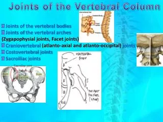
VertebralHémangioma (VH) • The most common benign tumor of the spine• Multiple in 25% of cases• Peak incidence: 40-60 years• Slow-growing lesion• benign vascular dysplasia capillary Cavernous (most common) or Venous INTRODUCTION (Picture takenfromwebsite:www.back.com/causes-tumors-benign.html)
When to treat a spinal hemangioma? Usually asymptomatic, discovered incidentally. Only 0.9% to 1.2% of cases become symptomatic:Aggressive Hemangioma Local pain, Radiological aggressiveness Neurologic deficit INTRODUCTION
Background: Semiology of vertebral HemangiomaRadiographic findings Vertical striations and trabeculations “Honeycomb” appearence. CT axial image “Polka dot” appearance of the involved vertebra MRI increased signal on T1- and T2 weighted images (intralesional fat)
Spine level between T3 to T10• Involvement of the entire vertebral body• Extension to the posterior arch• Discontinuous cortical bone • Lyticappearence• Paraspinal or intra ductal expansion• Low signal intensity on T1-weighted images • Intense enhancement after contrast injection Signs of aggressiveness on imaging of Vertebral Hemangioma
CASE N°1 A 18 years old patient 09/08/2007 Neurological dysfunction due to spinal cord compression.Radiographic findings: aggressive vertebral hemangioma T310/08/2007bilateral T3 laminectomyFollow-up: worsening paraparesisImmediate revision surgery: epidural hematoma evacuation
Significant improvement of motor deficit. • Histologic diagnose: capillary hemangioma
April 2009 (20 months later) • High back pain • Spastic paraparesis • Bilateral Babinski signs
MRI sequences a,b,csagittale T2-weighted images d : sagittale T1 weighted images withcontrast injection e : axial T1 weighted image withcontrast injection a b c d e Is there an explanation for the current neurological symptoms ?
What could be proposed?A. Reoperation B. TransarterialEmbolization C. Surgery with preoperative embolization D. vertebroplasty E. Radiotherapy
What could be proposed?A. Reoperation B. TransarterialEmbolization • C. Surgery with preoperative embolization D. vertebroplasty E. Radiotherapy
What arterial branches to explore?A. The celiac trunk and superior mesenteric artery B. The dorsal intercostal arteriesC. The lumbar arteriesD. The thoracic and abdominal aorta
What arterial branches to explore? • The celiac trunk and superior mesenteric artery • B. The dorsal intercostal arteries C. The lumbar arteries D. The thoracic and abdominal aorta
Which embolic agent to use ? A. Coils B. Embospheres C. Curaspon D. Ethanol E. Biological Glue
Which embolic agent to use ? A. Coils B. Embospheres C. Curaspon D. Ethanol E. Biological Glue
Which embolic agent to use ? A. Coils B. Embospheres C. Curaspon D. Ethanol E. Biological Glue
Which embolic agent to use ? A. Coils B. Embospheres C. Curaspon D. Ethanol E. Biological Glue
Which embolic agent to use ? A. Coils B. Embospheres C. Curaspon D. Ethanol E. Biological Glue
Which embolic agent to use ? A. Coils • B. Embospheres C. Curaspon D. Ethanol E. Biological Glue
The anterior spinal artery was identified in T10 left. Is there a risk of embolization of T3.A. yesB. noC. Distrust
The anterior spinal artery was identified in T10 left. Is there a risk of embolization of T3. • A. Yes B. No • C. Distrust
Embolization Right T4 Right T5
Selective angiography of the pedicle of the left T3intercostal artery We can embolize at this level? A. Yes B. No
Selective angiography of the pedicle of the left T3intercostal artery We can embolize at this level? A. Yes • B. No
Surgical resection is limited because of: A. The involvement of the anterior archB. The epidural extensionC. The involvement of the posterior arch
Surgical resection is limited because of: A. The involvement of the anterior arch • B. The epidural extension • C. The involvement of the posterior arch
What can we do ?A. Surgery as part of the angioma was embolizedB. VertebroplastyC. Sclerotherapy with Absolute ethanol D. There is no other treatmentE. There is another alternative ?
What can we do ?A. Surgery as part of the angioma was embolizedB. VertebroplastyC. Sclerotherapy with Absolute ethanol D. There is no other treatment • E. There is another alternative?
What does this alternative ?A. radiofrequency ablationB. direct embolization ?
What does this alternative ?A. Radiofrequency ablation • B.Direct embolization ?
Which embolic agent to use ? A. Ethanol B. Coils C. Embospheres D. Biological Glue
Which embolic agent to use ? A. Ethanol B. Coils C. Embospheres • D. Biological glue
Which type of radiographic guidance ? A. Fluoroscopy B. CT scanner C. Ultrasonography
Which type of radiographic guidance ? A. Fluoroscopy • B. CT scanner C. Ultrasonography
Sclerotherapy with Glubran 2 by direct puncture under CT guidance
Sclerotherapy with Glubran 2 by direct puncture under CT guidance
Sclerotherapy with Glubran 2 by direct puncture under CT guidance
Sclerotherapy with Glubran 2 by direct puncture under CT guidance
Sclerotherapy with Glubran 2 by direct puncture under CT guidance
Is surgery indicated ? A. Yes B. No
Is surgery indicated ? • A. Yes B. No
What time limits ? A. In 7 dayssothat the inflammation decreases B. In one month C. Within 48 hours of embolization D. The time limits is not important
What time limits ? A. In 7 dayssothat the inflammation decreases B. In one month • C. Within 48 hours of embolization D. The time limits is not important
Surgery should include : • T 3 Laminectomy • T 3 Vertebrectomy C. Laminectomy and osteosynthesis D. Osteosynthesis
Surgeryshouldinclude: A. T 3 Laminectomy B. T 3 Vertebrectomy • C. Laminectomy and osteosynthesis D. Osteosynthesis
Favorable evolution with recovery of motor function of lower extremities. • Is the treatmentachieved? • A . Yes • B . No
Favorable evolution with recovery of motor function of lower extremities. • Is the treatmentachieved? • A . Yes • B . No
To treat vertebral body of T3 must be associate : A. Surgery by anterior approachB. PercutaneousVertebroplastyC. Sclerotherapy with Glubran 2 under CT guidance