Download
1 / 41
410 likes | 415 Views
Evaluation of the Painful Shoulder. J. Lindsay Quade, MD Clinical Instructor Internal Medicine/Pediatrics, Sports Medicine University of Michigan Health System. Objectives. To improve physician comfort with obtaining relevant history in the evaluation of the painful shoulder

E N D
Evaluation of the Painful Shoulder J. Lindsay Quade, MD Clinical Instructor Internal Medicine/Pediatrics, Sports Medicine University of Michigan Health System
Objectives • To improve physician comfort with obtaining relevant history in the evaluation of the painful shoulder • To improve physician comfort with physical examination of the shoulder, including special testing • To improve physician comfort with diagnosis and management of common causes of shoulder pain
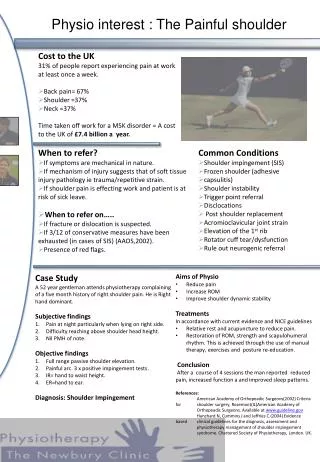
The Shoulder • Shoulder pain is common in the primary care setting, responsible for 16% of all musculoskeletal complaints. • Taking a good history, paying special attention to the age of the patient and location of the pain, can help tailor the physical exam and narrow the diagnosis. • Knowledge of common shoulder disorders is important as they can often be treated with conservative measures and without referral to a subspecialist.
MSK Shoulder Pain Differential • Articular Causes • Glenohumeral (GH) and acromoclavicular (AC) arthritis • Ligamentous and labral lesions • GH and AC joint instability • Osseus: fracture, osteonecrosis, neoplasm, infection • PeriarticularCauses • Chronic impingement and rotator cuff tendinitis • Bicep tendinitis • Rotator cuff and long biceps tendon tears • Subacromial bursitis • Adhesive capsulitis
Taking Your History • Age • Hand dominance • Occupation • Sports/physical activities • Trauma • Onset • Location • Character • Duration • Radiation • Aggravating/relieving factors • Night pain • Effect on shoulder function • Stiffness/restriction of movement • Grinding or clicking • Weakness • Numbness/tingling • Pain
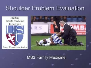
The Physical Exam • Inspection • Asymmetry • Bony deformity or abnormal contour • Muscle atrophy or bulge • Scapular winging
The Physical Exam • Range of Motion • Active • Passive • Apley’s “scratch” test • Scapular movement • Strength Testing
The Rotator Cuff Muscles • Supraspinatus • Abduction • Infraspinatus • External rotation • Subscapularis • Internal rotation • Teres minor • External rotation
The Physical Exam • Palpation • AC, SC, and GH joints • Biceps tendon • Coracoid process • Acromion • Scapula
Special Tests • Rotator Cuff • “Drop-arm” • “Empty can,” push-off, and resistance testing • Impingement • Neer’s • Hawkins
Special Tests • Biceps • Speed’s • Yergason’s • AC Joint • Cross-arm
Special Tests • Shoulder Instability • Sulcus sign • Apprehension, relocation, release • Load and shift
Special Tests • Labrum • O’Brien’s • Crank test • SLAPprehension
Specific Examples • Rotator Cuff Pathology • “Frozen Shoulder” • Shoulder Instability • AC Joint Separation • Arthritis • Labral Tear • “SICK Scapula” Main points: Presenting symptoms PE findings Diagnosis Conservative treatment or refer?
Rotator Cuff Pathology • Presentation & symptoms: • PAIN • +/- weakness • Age? trauma vs chronic • Physical exam findings: • Pain with ROM & resistance testing (+empty can, +push-off) • + drop arm if full-thickness tear • + Neer’s and Hawkins if impingement
Rotator Cuff Pathology • Diagnosis: • Xray – often negative • Ultrasound • Consider MRI if planning for surgery • Management: • Tendinopathy or impingement – conservative treatment, PT, subacromial GC injection • Partial-thickness tear – PT (up to 12 weeks), possibly subacromial GC injection • Full-thickness tear – Ortho referral
“Frozen Shoulder” (Adhesive Capsulitis) • Presentation & symptoms: • Pain, often >3 months • Progressive loss of ROM • Age >40yo • Risk factors: immobility, DM, hypothyroidism • Physical exam findings: • Limited active ROM, external rotation often 50% normal • Endpoint with passive ROM
“Frozen Shoulder” (Adhesive Capsulitis) • Diagnosis: • CLINICAL! • Xray if need to rule-out fracture or OA • US later if concerned for RC pathology • Management: • Set expectations • Pain control, gentle ROM exercises/PT • If severe, intra-articular GC injection under fluoroscopy
Shoulder (GH) Instability • Presentation & symptoms: • Pain • Instability • Age < 40yo • Transient neurologic symptoms • History of dislocation or subluxation • Physical exam findings: • + sulcus • + apprehension & relocation • + load & shift testing
Shoulder (GH) Instability • Diagnosis: • Clinical • Xrays often normal • MR arthrogram if no improvement • Management: • Activity modification • PT focused on aggressive strengthening • Refer to Ortho if no improvement with PT or if recurrent dislocation
Acute Shoulder Dislocation • Physical Exam: • External rotation & abduction, palpable humeral head • Check innervation of skin over lateral deltoid! (Axillary nerve) • Diagnosis: • Clinical • Xray • Management: • Relocate & immobilize • ROM exercises within 7-10 days aggressive rehab program
AC Joint Separation • Presentation & symptoms: • Direct blow to shoulder or FOOSH • Male contact sport athlete, ~20yo • Pain/swelling • Physical exam findings: • Pain and swelling over AC joint • “Stepped” deformity if more severe • + cross-arm test • + painful arc
AC Joint Separation • Diagnosis: • Clinical + • Xray • Management: • Types I-III: Non-operative (rest, ice, analgesics, sling for immobilization, PT) • Types IV+: Ortho referral for surgery
Shoulder Arthritis • Presentation & symptoms: • Age >50 • Progressive pain with activity • Decreased ROM • Impingement symptoms • History of rotator cuff injury, previous trauma, or shoulder surgery • Physical exam findings: • AC joint: tenderness over AC joint, pain at extreme internal rotation, + cross-arm test • GH joint: decreased ROM, pain and crepitus at extremes of motion
Shoulder Arthritis • Diagnosis: • Clinical + • Xray • Management: • AC joint: • Activity modification, NSAIDs, GC injection • GH joint: • Goal = maintain function with adequate pain control • PT, glucosamine & chondroitin, intra-articular GC injection • Referral to Ortho if conservative treatment fails
Labral Tear • Presentation & Symptoms: • Pain +/- instability • Clicking/popping • Overhead athlete, history of dislocation, history of trauma • Physical Exam: • Pain with passive external rotation • Pain with palpation of bicipital groove • + Apprehension/relocation, O’Brien’s, SLAPprehension, crank tests • Can also have + biceps testing
Labral Tear • Diagnosis: • Xrays usually normal but may show Hills-Sach lesion • MRI or MR arthrogram • Management: • Conservative: rest, NSAIDs PT • Operative: Ortho referral • If conservative measures fail, larger tears, concomitant RC tear
“SICK Scapula” • Presentation & Symptoms: • Pain • Repetitive overhead activity • Drooping shoulder on dominant side • Physical Exam: • Scapular malposition • Inferior medial border prominence • Coracoid pain and malposition • Kinesis abnormalities of scapula
“SICK Scapula” • Diagnosis: • Clinical • Management: • Physical Therapy & kinetic-chain based rehabilitation • Pain free ROM Strengthening Proprioception exercises
Take-Home Points • Shoulder pain is common • Taking a good history can help narrow both your differential and your physical exam • Having a good grasp of shoulder anatomy is necessary for interpreting physical exam findings • Develop your own shoulder exam approach and follow it consistently • Aim to know at least one special test for each category of shoulder pain
Take-Home Points • Rotator cuff pathology can often be diagnosed with US • Frozen shoulder is a clinical diagnosis • To be diagnosed with arthritis, there should be pain on exam and an abnormal xray • If you are concerned about a labral tear, consider referral +/- an MR arthrogram • Most chronic shoulder pain can be treated conservatively • If patient is not improving clinically, refer to Sports Medicine
YouTube! • “Complete Musculoskeletal Exam of the Shoulder” • by University of Michigan Family Medicine
References • Beuerlein MJS, McKee MD, Fam, AG. (2010). The shoulder. In Lawry GV (2nd.), Fam’s musculoskeletal examination and joint injection techniques, (pp. 7-19). Philadelphia: Mosby. • Burbank KM, Stevenson JH, Czarnecki GR, Dorfman J. Chronic shoulder pain: part I. Evaluation and diagnosis. Am Fam Physician. 2008 Feb 15; 77 (4): 453-60. • Burbank KM, Stevenson JH, Czarnecki GR, Dorfman J. Chronic shoulder pain: part II. Treatment. Am Fam Physician. 2008 Feb 15; 77 (4): 493-7. • Dodson CC, Altchek DW. SLAP lesions: an update on recognition and treatment. J Orthop Sports Phys Ther. 2009 Feb; 39 (2): 71-80. • Edmonds EW, Denerink DD. Common conditions in the overhead athlete. Am Fam Physician. 2014 Apr 1; 89 (7): 537-41. • Ewald, A. Adhesive capsulitis: a review. Am Fam Physician. 2011 Feb 15; 83 (4): 417-22. • O’Connor, F, et al. (2013). ACSM’s Sports Medicine: A comprehensive review. Musculoskeletal injuries in the tennis player, (pp. 717). Philadelphia: Lippincott. • Woodward TW, Best TM. The painful shoulder: part I. Clinical evaluation. Am Fam Physician. 2000 May 15; 61 (10): 3079-88. • Woodward TW, Best TM. The painful shoulder: part II. Acute and chr onic disorders. Am Fam Physician. 2000 Jun 1; 61 (11): 3291-300.
Photo References • 1. Slide 4: http://www.chiro.org/LINKS/Shoulder.shtml • 2. Slide 5: http://www.physiodc.com/shoulder-pain-with-yoga-adjust-your-downward-dog/ • 3. Slide 6: http://www.njorthoclinic.com/need-biceps-tenodesis-labrum-tear/ • 4. Slide 7: https://acewebcontent.azureedge.net/blogs/blog-examprep-091313-2.jpg • 5. Slide 10: http://www.pic2fly.com/Biceps+Popeye+Deformity.html ; http://www.wheelessonline.com/userfiles/2010-07-19%2015_44_46.jpg • 6. Slide 11: https://www.studyblue.com/notes/note/n/scapula--deltoid-regions/deck/3234274 (large picture); http://www.masmusculo.com.es/workout/el-apleys-scratch-test/ ; https://acewebcontent.azureedge.net/blogs/blog-examprep-091313.jpg (scapula pic) • 7. Slide 12: http://jimmysmithtraining.com/six-pack-diet/good-hurt-bad-hurt (left); http://createperformance.blogspot.com/2012/09/thanks-to-g.html • 8. Slide 13: http://www.physiodc.com/shoulder-pain-with-yoga-adjust-your-downward-dog/ • 9. Slide 14: http://meded.ucsd.edu/clinicalmed/joints2.htm (top picture); http://chrisjohnsonpt.com/are-you-taking-your-shirt-off-properly-2/
Photo References • 10. Slide 15: http://www.dynamicchiropractic.com/mpacms/dc/article.php?id=52373 (left picture); http://openi.nlm.nih.gov/detailedresult.php?img=2684214_12178_2008_9024_Fig4_HTML&req=4 • 11. Slide 16: http://www.arthritisresearch.us/rheumatoid-arthritis/the-shoulder.html (cartoon); https://www.youtube.com/watch?v=0_Y8twcQ9Ho • 12. Slide 17: http://ameblo.jp/g-money0229/entry-11512342937.html (cartoon); https://www.youtube.com/watch?v=3G6mb1QQ90I • 13. Slide 20: http://radiopaedia.org/images/1983198 • 14. Slide 21: http://www.myerssportsmedicine.com/frozen-shoulder/ • 15. Slide 23: http://shawchiroandsport.com/shoulder-joint-increased-mobility-means-increased-risk-injury/hypermobile-shoulders • 16. Slide 25: http://www.physiownc.com/shoulder-pain/ • 17. Slide 26: https://www.studyblue.com/notes/note/n/unit-17-exam-2/deck/6899151 • 18. Slide 27: http://quoteimg.com/ac-joint-separation-classification/
Photo References • 19. Slide 28: http://grutter.us/ShoulderArm/AC%20arthritis.html • 20. Slide 29: http://www.orthoinfo.org/topic.cfm?topic=A00222 • 21. Slide 30: http://josephhechtmd.com/slap-legion-surgery/ • 22. Slide 31: http://www.appliedradiology.com/articles/mr-arthrography • 23. Slide 32: http://www.sportsandortho.com/UserFiles/sick2.png • 24. Slide 33: https://acewebcontent.azureedge.net/blogs/blog-examprep-091313-2.jpg • 25. Slide 37: http://galleryhip.com/michigan-football-logo-go-blue.html • All photos obtained through Yahoo! or Google image search