Download

1 / 56
620 likes | 890 Views
Uveal Disease. Wen Xu. What are these?. Uveitis. Description:. Definition:

E N D
Uveal Disease Wen Xu
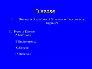
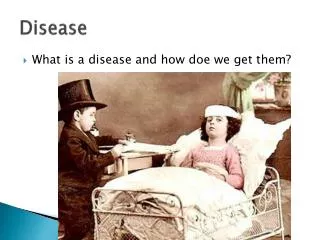
Description: • Definition: It denotes inflammation of the choroid(choroiditis),ciliary body(intermediate uveitis,peripheral uveitis,or pars planitis),or iris(iritis).However,common usage includes inflammation of the retina(retinitis), retinal vasculature(retinalvasculitis),and optic nerve(opticneuritis) • Features: It usually affects people 20-50 years of age and account for 10-15% of cases of legal blindness in developed countries. It is often combined with autoimmune diseases and characterised by its severe complications and recurrency
etiology and pathogenesis: (1)Externalcauses: infectiousdisorders (global penetration,intraocular foreign body, intraocular surgery and so on),noninfectious disorders (chemical burns,thermal burns,mechanical trauma or toxic stimulus). (2)Secondarycauses: secondary to inflammation of the global itself or adjacent tissues or toxic stimulus from intraocular disorders. (3)Internalcauses: infectious disorders (bacteria, viruses, fungi,parasites,protozoas),noninfectious disorders (combined with immunal or systemic disorders such as lens-induced uveitis,sympathetic ophthalmia and Behcet’s disease) (4)Inflammatorymediators: prostaglandins(PGS,PGE) (5)Oxidative damages(from free radical reactions)
classification: (1)Location-classified: anterior uveitis,intermediate uveitis,posterior uveitis and diffused uveitis (2)Clinical features: suppurative uveitis andnonsuppurative uveitis (3)Pathology: granulomatous uveitis and nongranulomatous (4)Etiology: infectious uveitis and noninfectious uveitis
Description: • It consists of iritis and iridocyclitis, often associated with ankylosing spondylitis or Reiter’s syndrome and so on • It is more common than any other type of uveitis and is usually unilateral and acute in onset
Clinical findings • Symptoms: pain,photophobia,tearing and blurred vision. • Signs: (1)ciliary or mixed congestion (2)keratic precipitates, KP (3)aqueous flare (4)iris changes (5)pupil changes (6)lens changes (7)vitreous and fundus changes
Ciliary or mixed congestion Episcleral vascular congestion around limbus.
Keratic precipitates,KP dust-like KP Middle-sized KP mutton fat KP
Aqueous flare Aqueous cells Hypopyon
Iris changes • Posterior synechia of iris • Iris bombe • Anterior synechia of iris • nodules • goniosynechia
Pupil changes • miosis • irregular shape • pupillary closure • occlusion of pupil
Lens changes Residual pigment on the surface of lens when the closed pupil opens again.
Vitreous and Fundus changes Cystoid macular edema or papilloedema cannot be seen frequently,but severe vision damage may occur once they happens.
Complications and Sequelae • Complicated cataract • Secondary glaucoma • Ocular hypotension • Atrophy of eyeball
Diagnosis 1.Typical clinical findings: symptoms and signs 2.Systemic disorders history: joint disease like juvenile rheumatoid arthritis and ankylosing spondylitis,Fuch’s heterochromic iridocyclitis,lens-induced uveitis and etc. 3.Laboratory testings: blood sedimentation accelerates, HLA-B27 histocompatibility antigen test (+), specific pathogen and etc.
Differential Diagnosis PACG acute iridocyclitis acute conjunctivitis Symptoms severe eye pain slight eye pain foreign body sensation headache photophobia burning nausea、vomiting tearing mucus or pus-like discharge Vision Markedly blurredSlightly blurred No effect on vision Congestionmixed ciliary or mixed conjunctival Cornea steamy edema、opacity transparent normal pigmentary KPhoar KP Pupil dilated and fixedmiosis normal vertical oval irregular shapes Anterior shallow、aqueousnormal or deepnormal Chamber slight opacity aqueous opacity IOP Markedly elevatednormal normal Treatment myotica mydriatic agents anti-inflammatories lower IOP anti-inflammatories antivirals
Treatment • Principles:dilate the pupil immediately in case of posterior synechia of iris;use anti-inflammatories rapidly to avoid eye tissue damage and complication occurrence; eliminate the pathogen. • Seldomly do: antibiotics and systemic medicines.
Plans: 1.cycloplegics:①prevent and seperate the posterior synechia of iris in case the complications occur. ② reduce discomfort from ciliary spasm. The first line is Homatropine drops or ointments; when severe inflammation occurs,our first choice comes to 1% Atropine, then change into Homatropine or tropicamide. 2.corticosteroids:Care should be taken to rule out an corneal epithelial defect in case of infection. Short-term systemic medicines and periocular injections areallowed when there is papilledema or macular edema.
3.nonsteroidal anti-inflammatory drugs (NSAIDs):aspirin or local drops4.antimicrobials:when caused by infection. 5.other immunosuppressants 6.treat the accompanied systemic disorders. 7.pathogen treatment. 8.other therapies:foment,and etc. 9.treat the complications and sequelae:anti-glaucoma surgeries,cataract surgeries under good control of the imflammation.
Intermediate uveitis, Peripheral uveitis
Description: • It consists of pars planitis、the imflammation of vitreous base and peripheral retina • It is the second most common type of intraocular imflammation,the hallmark of which is vitreous inflammation • It is typically bilateral、 slow in onset and tends to affect patients in their late teens or early adult years. Men are affected as commonly as women • The cause is unknown and autoimmune disorders are always considered
Clinical findings • Symptoms: floaters and blurred vision. Pain,photophobia and redness are usually absent or minimal • Signs: (1)anteriorsegment is general normal, but if significant, there may exist the manifestations of anterior uveitis like KP, aqueous flare, aqueous imflammation cells, posterior synechia of iris and etc.
(2) The most striking finding on examination is vitritis, often accompanied by vitreous condensates, either free-floating as “snowballs” or layered over the pars plana and ciliary body as “snowbanking” (3) There may exist macular edema and optic neuritis、peripheral retina vasculitis、vacular white sheath and etc. (4) Systemic disorders: Mutiple sclerosis, infection,Behcet’s disease,imflammatory bowel disease and etc.
Complications: (1) complicated cataract (2) secondary glaucoma (3) macular edema (4)macular degeneration (5)retinal or choroidal detachment
Diagnosis • Typical clinical findings: symptoms and signs • Systemic disorders • Ancillary tests: slit lamp with three-mirror lens、fundus fluorescein angiography
Treatment • Corticosteroids: drops、sub-Tenon’s sac injection or take orally • Other immunosuppressants: CsA. Pay attention to the toxicity and side-effects • Laser coagulation or cryocoagulation • Vitreous surgeries • Pathogen therapy
Description • Choroiditis • Choroidoretinitis • Retinochoroiditis • Neurochoridoretinitis • They may occur alone or combination
Clinical findings • Symptoms typically include floaters, loss of visual field or scotomas, or decreased vision, which can be severe • Retinal detachment, though infrequent, occurs most commonly in posterior uveitis and may be tractional, rhegmatogenous, or exudative in nature
Diagnosis and treatment • Diagnosis: typical vitreous、retinal and/ or choroidal diseases; systemic disorders;fundus fluorescein angiography or ICGA;laboratory tests and other ancillary tests to determine the cause or type • Therapy: anti-infectious treatment; corticosteroids;other immunosuppressants; surgeries
Description • The term “diffuse uveitis” is used to denote a more or less uniform cellular infiltration of both the anterior and posterior segments. Associated findings such as retinitis、vasculitis、or choroiditis can occur and often prompt further diagnostic testing • Tuberculosis,sarcoidosis, and syphilis should always be considered in patients with diffuse uveitis • Vogt-Koyanagi-Harada syndrome and Behcet’s disease are the most common types
Diagnosis: contain both the manifestations of anterior and posterior uveitis Treatment: do as anterior and posterior uveitis
Vogt-Koyanagi-Harada syndrome • It is a typically bilateral,granulomatous, recurren,diffuse uveitis combined by systemic meningismus,hearing impairment, vitiligo,whiten or fallen hair • Ocular manifestations: decreased vision, sunglow-like fundus,Dalen-Fuchsnodules, complicated cataract, secondary glaucoma and etc.
Diagnosis: according to history, clinical manifestations,fundus fluorescein angiography, ncurolympy test Treatment: corticosteroids,other immunosuppressants,surgeries
Behcet’s disease • It is a marked by recurrent diffuse uveitis,recurrent canker sore,polymorphous skin lesions and genital ulcer, involved in multiple systems, thus becomes a stubborn disease • Diagnosis and treatment: according to clinical manifestations and laboratory tests results; therapy includes mydriatic agents, corticosteroids, immunosuppressants and surgeries under good control of imflammation
Fuchs heterochromic uveitis • It is a chronic nongranulomatous uveitis, marked by iris hopochromia or atrophy. Anterior uveitis is the most common type • Patients usually complain of blurred vision. KPs are often small and stellate and scattered over the entire endothelium.Telangiectatic blood vessels may be seen in the chamber angle on gonioscopy.Possible complications are subcapsular cataract and higher IOP • Therapy: anti-inflammatories, lowing the IOP or surgeries
Acute retinal necrosis syndrome; ARN • It is a severe unilateral diffuse uveitis accompanied by retinal arteritis,retinal nacrosis,severe vitreous opacity and subsequent retinal detachment • Herpes simplex and herpes zoster are the most common causes. Adults are more susceptible. No sex difference. It is intractable to treat and prognosis is bad
Diagnosis: clinical manifestations,laboratory tests, PCR, biopsy Treatment: antivirals(Gancyclovir, Acyclovir), anticoagulants (heparin,small doses of aspirin), corticosteroids, laser coagulation or surgeries
Tumors Involving the Uveal Tract • Malignant melanoma of the choroid • Choroidal hemangioma • Metastatic carcinoma of choroids • Iris cyst
Malignant melanoma of the choroid • It is the most commonintraocular malignant tumor in the white population, second to retinoblastoma in China. It comes from pigment cells and nevus cells • It may be seen in its early stages only accidentally during routine ophthalmoscopic examination or because of blurring due to macular invasion. Blood-borne metastases may occur at any time. Glaucoma may be a late manifestation
Intraocular malignant melanomas mayextentinto adjacent intraocular tissues or outside the eye through the scleral canals or by intravascularinvasion • Clinical manifestations are usually absent unless the macula is involved. In the later stages,grows of the tumor may lead toretinal detachmentwithdecreased visionandloss of visual field • Dome-shaped or mushroom-like tumor can be seen ophthalmoscopically
Four stages:intraocular stage,glaucoma stage,extraocular extension stage and systemic metastasis stage • Diagnosis tips: ①early diagnosis is difficult. ②typical fundus changes and symptoms. ③FFA,ICGA,ultrasonic,MRI,CT and biopsy may be of some value • Treatment:local coagulation, local cryocoagulation,TTT,radiotheraphy,local resection and enucleation of eyeball