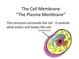
Download
1 / 8
150 likes | 603 Views
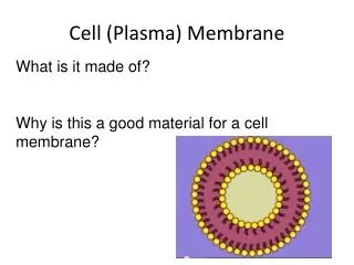
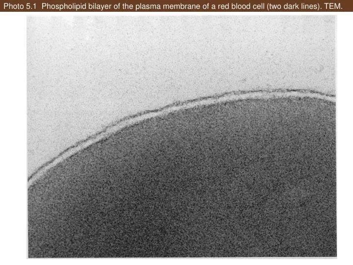
Photo 5.1 Phospholipid bilayer of the plasma membrane of a red blood cell (two dark lines). TEM. Photo 5.2 Proteinaceous particles in the plasma membrane of Rhodospirillum rubrum . TEM. Photo 5.3 Freeze-fractured, freeze-etched cell of cyanobacterium ( Synechococcus lividus ). TEM.

E N D
Photo 5.1 Phospholipid bilayer of the plasma membrane of a red blood cell (two dark lines). TEM.
Photo 5.2 Proteinaceous particles in the plasma membrane of Rhodospirillum rubrum. TEM.
Photo 5.3 Freeze-fractured, freeze-etched cell of cyanobacterium (Synechococcus lividus). TEM.
Photo 5.8 Plasmolyzed cells in Anacharis (Elodea) sp. leaf in salt solution. LM.
Photo 5.9 Plasmolyzed cell of Escherichia coli B, caught in the process of dividing (fission). TEM.
Photo 5.10 Lignified secondary cell walls with numerous plasmodesmata in Lunaria annua. LM.
Photo 5.11 Fertilization causes a wave of calcium ions to pass through the egg of a sea star.