Download
1 / 18
180 likes | 182 Views
This article provides an overview of the primary functions of the respiratory system, including respiration, inspiration, and expiration. It also discusses the various structures of the respiratory system, such as the nose, nasal cavity, pharynx, larynx, trachea, bronchi, and lungs.

E N D
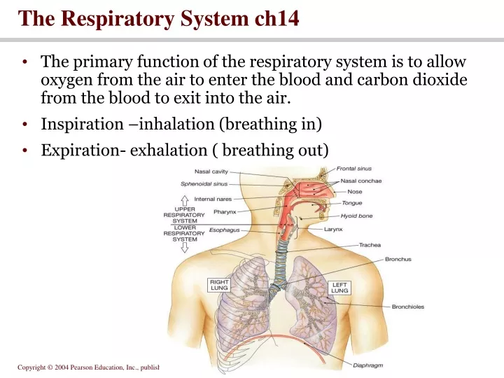
The Respiratory System ch14 • The primary function of the respiratory system is to allow oxygen from the air to enter the blood and carbon dioxide from the blood to exit into the air. • Inspiration –inhalation (breathing in) • Expiration- exhalation ( breathing out)
The Nose • Functions • Provides an airway for respiration • Moistens and warms entering air • Filters and cleans inspired air • Resonating chamber for speech • Detects odors in the airstream • Respiratory mucosa-contains goblet cells that secrete mucus • Mucus • Stickiness traps inhaled particles • Lysozyme kills bacteria
Nasal cavity • Vibrissae (guard hairs) – stiff hairs that filter large particles from the air • Nasal cilia – hair-like projections that propel trapped particles towards the throat for digestion by digestive enzymes
Rich supply of capillaries warm the inspired air • Nasal conchae – folds in the mucous membrane that increase air turbulence and ensures that most air contacts the mucous membranes • Olfactory mucosa – mucous membranes that contain smell receptors
The Nose, Nasal Cavity, and Pharynx Figure 23.3c
The Pharynx (throat) • Funnel shaped passageway that connects the nasal and oral cavities to the larynx • Three regions of the pharynx • Nasopharynx – air passage • Oropharynx – passageway for air, food, and drink • Laryngopharynx – passageway for air, food, and drink
The Larynx (voice box) • Functions : • Keeps food and drink out of the airway • Sound production • Anatomical Features: • Nine c-rings of hyaline cartilage form the framework of the larynx (the apex of this triangular box is called the Adam’ss apple • Muscular walls aid in voice production and the swallowing reflex • Glottis – the superior opening of the larynx • Epiglottis – prevents food and drink from entering airway when swallowing • False vocal cords – aid in closing the glottis when swallowing • True vocal cords – produce sound when air passes between them
The Anatomy of the Larynx Figure 23.4
The Glottis Figure 23.5a, b
The Trachea (windpipe) • Functions : • Air passageway • Cleans, warms, and moistens incoming air • Anatomical features : • Rings of hyaline cartilage – reinforce the trachea and keep it from collapsing when you inhale • Traps inhaled debris and propels mucus up to the pharynx where it is swallowed
The Anatomy of the Trachea Figure 23.6a, b
Bronchi • Function : • Solely an air passageway • Anatomical features : • Left and right primary bronchi branch off from trachea • Once the left and right primary bronchi enter the lungs they are subdivided into smaller tubes: Secondary bronchi (one for each lobe) → tertiary bronchi → bronchioles → terminal bronchioles → respiratory bronchioles → alveolar ducts → alveolar sacs
The Bronchi and Lobules of the Lung Figure 23.10a
The Lungs • Left • Divided into 2 lobes • Smaller than the right lung • Cardiac notch accommodates the heart • Right • Divided into 3 lobes • Each lobe is separated by connective tissue and has its own arteries and veins. • Serous membranes-cover the entire surface of the lungs and produce pleural fluid-enables the lungs to expand and contract with minimal friction • Visceral –adheres to the surface of the lung • Parietal- lines the thoracic cavity
The Gross Anatomy of the Lungs Figure 23.7
The Bronchi and Lobules of the Lung Figure 23.10b
The Alveoli • Alveolar sacs-clusters of alveoli • Alveoli- the site of gas exchange which occurs between the air in the alveoli and capillaries • Alveolar cells – allow for diffusion of gases & secrete surfactant- reduces the surface tension of fluid in the lungs and helps make (alveoli) more stable. This keeps them from collapsing when an individual exhales
Alveolar Organization Figure 23.12a-c