Download
1 / 11
110 likes | 218 Views
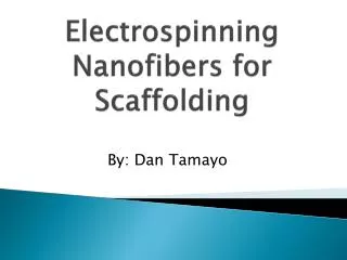
Chondrogenic Differentiation of hMSCs on PCL Nanofibers. Winnie Kuo University of California, Berkeley Final Presentation for NSF-REU at UIC August 3, 2006 Advisors: Prof. Cho, Prof. Megaridis, Joel Wise. Background.

E N D
Chondrogenic Differentiation of hMSCs on PCL Nanofibers Winnie Kuo University of California, Berkeley Final Presentation for NSF-REU at UIC August 3, 2006 Advisors: Prof. Cho, Prof. Megaridis, Joel Wise
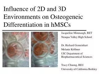
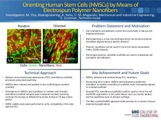
Background • Human Mesenchymal Stem cells (hMSCs) can differentiate into many cell lineages • Chondrogenesis -- cartilage repair therapy • Electrospun PCL nanofibrous scaffolds are biodegradable & mimic extracellular matrix Figure 1. hMSCs in culture Figure 2. Oriented nanofibers NSF REU at UIC Site
Goals • Mimic thin superficial layer of articular (joint cartilage) • Attach & Differentiate hMSCs into cartilage cells on polymer nanofiber scaffolds • Observe cell morphology & differentiation based on physical cues Figure 3. Articular cartilage NSF REU at UIC Site
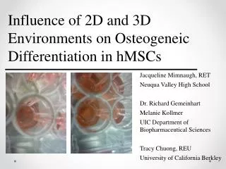
Project Design • Seed hMSCs on nanofibrous scaffolds • Cultured with chondrogenic media in 96-well plates (control with growth media) • Monitor cell proliferation & differentiation: • Fluorescence imaging • Total DNA count • Sulfated Glycosaminoglycan (sGAG) NSF REU at UIC Site
Cell Morphology Chondrogenic cells on nanofiber scaffold Chondrogenic cells on PCL film scaffold Mes. stem cells on nanofiber scaffold NSF REU at UIC Site
Tracking Differentiation Amount of sGAG* detected per μg of DNA *sGAG: cartilage-specific marker NSF REU at UIC Site
Conclusions • Cells on nanofibers proliferate in an oriented manner • Chondrogenic media and fiber alignment induce chondrogenesis • By 5th week, chondrogenic cells produced high amounts of sGAG • Oblong chondrogenic cell shape resembles superficial layer of articular cartilage NSF REU at UIC Site
Future Directions • hMSCs cultured on nanofibers as an alternative source of cartilage cells • Advantage: “renewable” • Incorporate cartilage-inducing factors within nanofibers • Chemicals & proteins contained within fibers may mimic ECM better than mere suspension NSF REU at UIC Site
Acknowledgements • NSF EEC-0453432 Grant • Novel Materials and Processing in Chemical and Biomedical Engineering (Director C.G. Takoudis) • Funded by the DoD-ASSURE and NSF-REU Programs • Professor M. Cho • Professor C. Megaridis • Professor A. Yarin • Joel Wise NSF REU at UIC Site
References • Reneker, D.H. et al, Electrospinning of Nanofibers from Polymer Solutions and Melts, Advances in Applied Mechanics, Vol. 41, Elsevier Inc. 2006. • Tuan, R.S., Song, L., Baksh, D., Adult mesenchymal stem cells: characterization, differentiation, and application in cell and gene therapy. J. Cell. Mol. Med. Vol 8, No 3: 301-316 2004. • Tuan, Boland, Tuli, Adult mesenchymal stem cells and cell-based tissue engineering. Arthritis Research & Therapy 2003, Vol 5: 32-45. • Calendar, R., The Three-Dimensional Structure of Proteins, Molecular & Cell Biology 102 Lecture Notes, Berkeley, CA, 2006. • Green, N., Wise, J., Cho, M., Megaridis, C., Quantitative analysis of human mesenchymal stem cell alignment by electrospun polymer nanofibrous scaffolds, University of Illinois, Chicago, 2005. • Li, W. et al, A three-dimensional nanofibrous scaffold for cartilage tissue engineering using human mesenchymal stem cells, Biomaterials, Vol. 26, Issue 6: 599-609, 2005. • http://en.wikipedia.org/wiki/Polycaprolactone, “Polycaprolactone” Wikipedia, the free encyclopedia. • http://ucalgary.ca/~kmuldrew/cryo_course/cryo_chap9_1.html, “Cryopreservation and Banking of Tissues” Ken Muldrew, 1999. • http://www.ukcte.org/gci.htm, “Cell Phenotype & Function” UK Centre for Tissue Engineering • http://www3.imperial.ac.uk/bioengineering/research/physiologicalfluidmechanics/transportintissue, “Transport in Tissue” Imperial College London, Dept. of Bioengineering NSF REU at UIC Site
Thank You! NSF REU at UIC Site