Download
1 / 40
400 likes | 488 Views
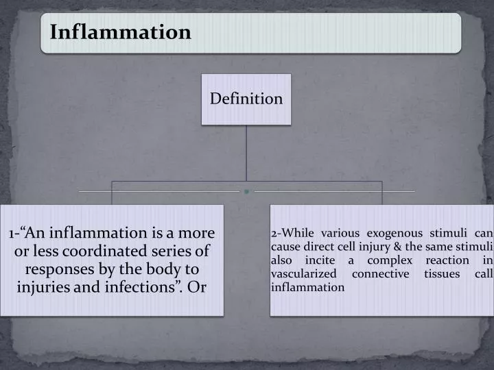
C:UsersDr Alok TripathiPicturesh_granulocytesPathway.gif. Diagrammatic Presentation . INCREASED. ECLIPASED. Permit. Functional mechanism is another mechanism resulting vascular permeability . Extravasation.

E N D
C:\Users\Dr Alok Tripathi\Pictures\h_granulocytesPathway.gif
INCREASED ECLIPASED Permit
Functional mechanism is another mechanism resulting vascular permeability
Extravasation Extravasation of leukocytes and lymphocytes, for example, seems to depend on a complex series of interactions between the membranes of the blood cells and the endothelial cells of the blood vessels. A variety of cell surface receptors, cell adhesion molecules, and secreted chemokines induce tight, specific interactions among the cell types, followed by migration between endothelial cells and out of the blood vessel. In addition to direct cell-cell interactions, there are also long range interactions that direct cells out of the blood stream and into the tissues at specific locations. These are mediated by chemokines (a special subclass of cytokines) that are released by a variety of lymphoid and myeloid cell types and that act through chemokine receptors to act as "chemo-attractants", luring cells along a concentration of chemokines to a particular location. All chemokine receptors known are G-protein coupled receptors that activate a variety of second messenger systems to mediate, among other things, directional cell movement. While the acute inflammatory response is an effective way to isolate an infection and to attract the macrophages, neutrophils, and lymphocytes that can attack and overcome it, under some conditions an inflammatory response can persist for long periods of time, becoming a chronic inflammatory response. This can occur because of an autoimmune disease (such as rheumatoid arthritis), because of persistence of an infection (such as tuberculosis), because of repeated tissue injuries ("tennis elbow"), or because of cancer. Often the chronic inflammation causes a mass of activated macrophages, lymphoctes, and antigen that form a tumor-like lump called a granuloma. Apparently one of the central activators of a chronic immune response is gamma-interferon, which is also essential for normal immune responses. Chronic inflammations are treated variously with drugs like aspirin and ibuprofen (non-steroidal anti-inflammatory drugs), steroids, or drugs that block extravasation. Presumably inhibitors of IFN-would also be effective, but I don't know of any that are available. The hottest news in anti-inflammatory drugs are the so called Cox (cyclooxygenase) inhibitors that are much more specific than other NSAIDs in blocking inflammation.
WEBSITE FOR ANNIMATION • http://www.gluegrant.org/flash/injury.swf • http://www.biostudio.com/index_%20inflammation%20mac.htm • http://library.med.utah.edu/WebPath/INFLHTML/INFL070.html
Inflammation Triggered by tissue damage due to infection, heat, wound, etc. Four Major Symptoms of Inflammation: 1. Redness 2. Pain 3. Heat 4. Swelling May also observe: 5. Loss of function
Functions of Inflammation 1. Destroy and remove pathogens 2. If destruction is not possible, to limit effects by confining the pathogen and its products. 3. Repair and replace tissue damaged by pathogen and its products.
Stages of Inflammation 1. Vasodilation: Increase in diameter of blood vessels. Triggered by chemicals released by damaged cells: histamine, kinins, prostaglandins, and leukotrienes. 2. Phagocyte Migration and Margination: Margination is the process in which phagocytes stick to lining of blood vessels. Diapedesis (Emigration): Phagocytes squeeze between endothelial cells of blood vessels and enter surrounding tissue.
Stages of Inflammation (Continued) Phagocytes are attracted to site of infection through chemotaxis. Phagocytes destroy microbes, as well as dead and damaged host cells. 3. Tissue Repair: Dead and damaged cells are replaced.
Antimicrobial Substances: I. Complement System: Large group of serum proteins that participate in the lysis of foreign cells, inflammation, and phagocytosis. Two mechanisms of complement activation: 1. Classical Pathway: Initiated by an immune reaction of antibodies. 2. Alternative Pathway: Initiated by direct interaction of complement proteins with microbialpolysaccharides. Both pathways cleave a complement protein called C3, which triggers a series of events.
Classical Complement Pathway is Triggered by Antibodies Binding to Foreign Cells
Both Classical and Alternative Complement Pathways Trigger the Cleavage of C3
Consequences of Complement Activation: 1. Cytolysis: Due to the formation of a membraneattackcomplex (MAC) which produces lesions in microbial membranes. 2. Inflammation: Complement components (C3a) trigger the release of histamine, which increases vascular permeability. 3. Opsonization: Complement components (C3b) bind to microbial surface and promote phagocytosis. 4. Inactivation of Complement: Regulatory proteins limit damage to host cells that may be caused by complement.
Classical and Alternative Complement PathwaysCause Inflammation, Opsonization, and Cytolysis
II. Interferons: Antiviral proteins that interfere with viral multiplication. • Small proteins (15,000 to 30,000 kDa) • Heat stable and resistant to low pH • Important in acute and short term infections. • Have no effect on infected cells. • Host specific, but not virus specific. Interferon alpha and beta: Produced by virus infected cells and diffuse to neighboring cells. Cause uninfected cells to produce antiviral proteins (AVPs). Interferon gamma: Produced by lymphocytes. Causes neutrophils to kill bacteria.