Download

1 / 39
400 likes | 484 Views
Homeostasis Overview & The Kidney. Chapter 7. Introduction – 7.1. As we know our bodies are a collection of many cells, which have become specialized over many years. It is important however that all of these cells work in a concerted effort to ensure our health.

E N D
HomeostasisOverview & The Kidney Chapter 7
Introduction – 7.1 • As we know our bodies are a collection of many cells, which have become specialized over many years. It is important however that all of these cells work in a concerted effort to ensure our health. • Factors like: temperature, pH and concentrations of glucose and oxygen have to remain at a constant level so normal functions will continue in our bodies (especially those driven by enzymes). • This process is called homeostasis.
Negative Feedback • Beginning to end is a negative feedback loop and the goal is to resist change • Regulation of blood glucose levels is an example • after a meal, glucose in the blood is high • messages sent to the brain (coordinating centre) sends message to the liver (regulator or effector) • causes excess glucose to be stored as glycogen • it is important to realize that the reverse can also happen
Positive Feedback • Reinforce change system moves the controlled variable even further away from a steady state. • Positive feedback is much more rare than negative feedback. • Labor strong contraction in uterus is maintained by the hormone oxytocin (released from the hypothalamus). This contraction will continue as long as the fetus’s head is pushing against the uterus. The more pressure, the more oxytocin that gets released and this continues until the baby is expelled and therein the contractions stop, which stops the release of oxytocin.
Components of Homeostasis - 7.2 • Sensory receptors • These test the bodies temperature, pH, glucose levels and blood pressure • If there is a deviation a sensory receptor sends a signal to coordinating centre (an organ capable of bringing about change to internal conditions) • Hypothalamus – regulates temperature and amount of hormones to put into blood • Message then sent to an regulator, which is a specific tissue, or organ that changes its function in response to the coordinating centre’s message.
Components of Homeostasis - 7.2 • Example: body temperature rises (sensory receptors), hypothalamus (coordinating centre) signals blood vessels (regulator) to dilate thereby releasing heat out through the skin until correct temperature is attained (sensory receptors).
The Importance of Excreting Wastes - 7.3 • Nitrogenous wastes • Removal of amino groups from amino acid formation produces toxic ammonia (NH3) • 3 general solutions to this problem: • Flushing– freshwater and saltwater fish break down proteins in their gills so little ammonia actually enters the body • Detoxification – mammals and land animals link amino groups (in liver) to carbon dioxide forming harmless urea. This process requires energy (3ATP/1 urea). Urea is carried to kidneys, which excrete urea as a principle component of urine. • Insolubilization – birds and terrestrial reptiles have eggs, which are in shells therein, no excretion is possible. Ammonia is converted to uric acid, which is insoluble, which can be stored in crystal form (lengthy and energy consuming).
Excretion Development • Freshwater Fish • Kidneys evolved in these species first • Body is hypertonic to water in which it lives (water tends to enter the body) so water is not reabsorbed by the nephrons • excess water through nephrons (ammonia and other wastes added) bladder urine
Excretion Development • Marine Fish • evolved after freshwater fish (believed due to fossil records) • hypotonic to water in which they live (water tends to leave body) therefore they take a lot of water in and excrete a lot of salt (do not reabsorb) however reabsorb as much water as possible • kidneys therefore are different all ions are pumped out (Ca2+, Mg2+, SO42-, PO43-)
Excretion Development • Amphibians and Reptiles • Amphibians = the first terrestrial vertebrate • Identical kidney to freshwater fish due to their habitat (choice of water or land) • Reptiles = have different habitats therein a lot of different kidney systems • Freshwater – (some crocs and alligators) same as freshwater fish • Marine – (some crocs, turtles, lizards, snakes) same as freshwater but eliminate salt via salt glands located near nose or eye • Terrestrial – reabsorb as much H2O as possible. Urine is as concentrated as blood plasma, if more, water would start to travel in. Urine is released as solid urea or uric acid
Excretion Development • Mammals and Birds • can remove far more water then any other kidney type and this is a good thing because we don’t live in water. • Human urine is 4x more concentrated as blood plasma (can be 20x in rodents) • This is achieved by increasing local salt concentrations around nephron tubes and water is then reabsorbed into surrounding bloodstream • Mammalian Kidney – p.346 (basic) and p.347 (indepth) • Countercurrent flow is the key • Involves passage of two solutes across the membrane of the loop (salt and urea)
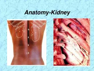
Urinary System – 7.4 • wastes filtered from the blood are transported to the urinary bladder via ureters. From the urinary bladder urine enters the urethra and is voided (600mL max). • Three main structures to the kidney • Cortex – outer connective tissue. surrounding part of an organism and in the kidney is the outer part (upper portion of the loop) • Medulla – area inside the cortex. inner or deep part and in the kidney it is the inner part (lower portion of the loop) • Renal pelvis – hollow chamber which joins the kidney and ureter.
The Nephron • About one million repeating structures called nephrons which are composed of the three factors: • Filter at the top of each nephron and is called Bowman’s capsule. In each capsule an arteriole enters and splits into a network of vessels called a glomerulus. Walls of capillaries act a filtration device, which gets unwanted particles out. • Tube Bowman’s capsule is attached to a long tube (renal tubule). There is a hairpin loop between the proximal and distal arms called the loop of Henle and it acts as an absorption point. • Duct the renal tubule empties into a large collecting duct, which aids as a water conservation device. Urine is 4 times as concentrated as blood plasma and a lot of water is taken out before urine is excreted. • This works because the duct is bent back alongside the renal tubule therefore there is salty tissue around and water will move out.
Formation on Urine – 7.5 • Filtration – glomerulus and Bowman’s capsule • The narrow walls of the glomerulus allows pressure to push small molecules into Bowman’s capsule. Large particles like proteins do not pass through. • Glomerular filtrate is the material that does pass through (water, nitrogenous wastes [urea], and nutrients [glucose and AA]) • Water passes out in the descending arm because the surrounding tissue has a high concentration of urea. • Loop of Henle is permeable to salts. There is a low salt concentration surrounding the Loop of Henle so salt (NaCl) passes out.
Formation on Urine • Reabsorption – proximal tubule, loop of Henle, distal tubule • Ascending arm utilizes active transport to pump ions out. This encourages even more water to travel out of the arm and be reabsorbed. • Without this reabsorption we would lose a lot of water and as such we would need to drink 1L of fluids every 10 minutes. • Collecting Duct is permeable to urea and it is passed out due to lower concentration outside the duct. This will allow water to travel out. It is this process of creating a high urea concentration that allows the water to pass out in step 1.
Formation on Urine • Secretion – colleting duct • Collecting Duct connects to others and forms the ureter, which transports wastes to the bladder and then out of the body via the urethra.
Water Balance – 7.6 • The Importance of Water Balance and Excretion • 67% of every vertebrate is water • we are continually taking in water but how do we get rid of it? • Respiration – expel water vapor and CO2 as we talk and breathe • Excretion – nitrogenous waste products, enzymes, acids, detergents, water, salts and ions
Water Balance – 7.6 • Salts and Ions • the blood’s osmotic concentration (concentration of solutes dissolved in it) must be kept at a steady state. • If you drink water the kidneys will remove excess water by increasing urine production • If sodium levels become too low the brain signals the adrenal gland to send out hormones that cause the kidneys to extract more sodium from the water passing through. • Conversely, if the sodium levels become too high the reverse will happen and more sodium will be allowed to pass through the kidneys. • The ratio or balance of salt to water is called osmoregulation
Osmoregulation • Maintaining a constant internal solute concentration, regardless of environment • Allows for complex patterns of internal metabolism, but also requires constant internal water regulation • Osmoconformers – vary osmotic concentration of body fluids to maintain an osmotic concentration that is close to that to of the medium in which they are living (marine invertebrates and sharks)
Osmoregulator examples • Freshwater vertebrates are hypertonic to surrounding water (high ion concentration inside so water tends to travel in) and will therein attempt to expel water. • Marine vertebrates are hypotonic relative to environment and water tends to leave their bodies so they will attempt to retain water. • Land animals need to retain all water possible to prevent dehydration
How does it work? • Idea is to maintain osmoregulation and this is achieved through an animal’s excretory system • Getting rid of wastes is coupled with water and ion concentration. What vertebrates do is push blood through a filtering system that gets rid of wastes but extracts all useful ions, nutrients and water back into the body.
Regulating ADH - Control of Water and Salt • ADH helps regulate the osmotic pressure of body fluids by causing the kidney to increase water reabsorption. • When ADH is released more concentrated urine is released • ADH is produced in the hypothalamus and stored in the pituitary gland
Regulating ADH - Control of Water and Salt • receptors allow recognition in hypothalamus of changes in osmotic pressure • decrease of water intake or increase water loss will cause blood solutes to become more concentrated increase the blood’s osmotic pressure! • consequently water will move into the bloodstream causing cells of the hypothalamus to shrink • signals release of ADH increases water reaborption and you produce more concentrated urine behavioural response you get thirsty as you drink you essentially do the opposite of above.
ADH and the Nephron • 85% of water filtered into the nephron is reabsorbed in the proximal tubule. • Decending loop of Henle is permeable to water and ions but ascending is permeable to NaCl. • ADH • Makes the upper part of the distal tubule and collecting duct permable to water • Large amount of NaCl in the intercellular spaces draws water from the upper section of the distal tubule and the collecting duct
Kidneys and Blood Pressure • Regulation of blood pressure by adjusting for blood volumes. • Aldosterone – acts on the nephrons to increase Na+ reabsorption. • Produced in the cortex of the adrenal gland, above the kidney. • As NaCl reaborption increases, the osmotic gradient increases and more water moves out of the nephron by osmosis.
pH Balance • pH of body 7.3-7.5 • Cellular respiration produces CO2 carbonic acid H+ ions lower pH • Buffer system of blood removes excess H+ ions; the buffer must be restored • the kidney helps to restore the buffer by reversing the reaction
pH Balance • CO2 is actively transported from the area around the nephron, into the cells that line the nephron. • CO2 combines with water to reverse the reaction (HCO3- and H+) • Bicarbonate ions diffuse back into the blood. • The H+ ions combine with phosphate ions or ammonia to be excreted.
Kidney Disease – 7.7 • Kidney Failure • Life threatening event as all fluids, which are vital, can be altered. • Infection, diabetes, high blood pressure • Urea and H+ collect in the blood if problems in the kidney
Kidney Disease – 7.7 • Diabetes Mellitus • caused by inadequate insulin from islet cells in the pancreas • blood sugar levels rise which remains in the nephron and inherently this extra solute throws off the osmotic balance of the nephron and water and solutes are voided in excess. • Diabetes Insipidus • destruction of ADH-hormone producing cells in the hypothalamus or the nerve tracts leading from the hypothalamus to the pituitary gland. • Urine output increases dramatically as there is no ADH to regulate water absorption
Kidney Disease – 7.7 • Bright’s Disease • Nephritis is inflammation of the nephrons caused by toxins from invading microbes (theory) • Proteins are able to pass into the nephron and consequently so does water to balance osmotic pressure. • Kidney Stones • mineral solutes from blood collect in the kidney (renal pelvis or ureter) • alkaline or acid stones • rip on the way out ouch!
Kidney Disease – 7.7 • Dialysis – removes toxic wastes from blood, utilizes a machine for a filtering mechanism. The main principles of a kidney are followed though… diffusion & blood pressure. There are two types: • Hemodialysis • tubes called catheters are surgically inserted into an artery and vein • catheters are attached to a dialysis machine every few days • use a thin cellulose acetate membrane to filter out unwanted materials • dialyzing fluid is the same as blood plasma but no unwanted materials are in it so diffusion will occur • need to carefully watch salt and water levels on own
Kidney Disease – 7.7 • Continuous Ambulatory Peritoneal Dialysis • patients own peritoneal membrane lining is used in the patient’s own abdominal cavity • catheter used to fill cavity with dialysis fluid and the body filters blood on its own but in a different region then normal (abdominal cavity)
Kidney Disease – 7.7 • Kidney Transplant • the other methods of dealing with kidney problems are band aid solutions but this method is a permanent solution. • The problem with this method is that cells are very specific. That is, there are self markers on the surface of all cells called histocompatibility antigens (fingerprints) and cells that might be introduced into another person’s body don’t have these so they will be attacked. • Only twins have the same markers so when a new kidney is introduced cyclosporin needs to be used, which suppresses the body’s immune system. The problem with this drug is that it allows an increase in infections (as immune system is lowered) and can be toxic to the liver and bone marrow.