Download
1 / 48
480 likes | 575 Views
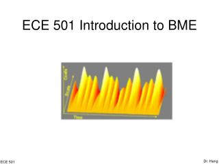
ECE 501 Introduction to BME. Dr. Hang. ECE 501. Part IV Bioinstrumentation Electrocardiogram. Dr. Hang. ECE 501. Introduction. Physiology Medicine Instrumentation. Dr. Hang. ECE 501. Medicine. The Electrical Cycle. Dr. Hang. ECE 501. Medicine. The Electrocardiogram.

E N D
ECE 501 Introduction to BME Dr. Hang ECE501
Part IV BioinstrumentationElectrocardiogram Dr. Hang ECE 501
Introduction • Physiology • Medicine • Instrumentation Dr. Hang ECE 501
Medicine • The Electrical Cycle Dr. Hang ECE 501
Medicine • The Electrocardiogram • The electrocardiogram (ECG) is a standardized • way to measure and display the electrical • activity of the heart. • • Physicians can diagnose problems with the • heart by analyzing its ECG and comparing it to • the ECG of a healthy heart. Dr. Hang ECE 501
Medicine • ECG Waves Dr. Hang ECE 501
Medicine • ECG Intervals Dr. Hang ECE 501
Medicine • A-V Blocks • Complete Hear Block • The cells in A-V node are dead and A-V node does not conduct action potential at all • Atria and ventricles beat independently • Ventricles are driven by abnormal pacemakers Dr. Hang ECE 501
Medicine • A-V Blocks • First-degree Hear Block • The A-V node is diseased • The action potentials from atria can still reach ventricles • It is greatly delayed Dr. Hang ECE 501
Medicine • Heart Flutter • Paroxysmal Tachycardia • An abnormal pacemaker in ventricle may discharge at a rapid rate • Ventricles depolarize irregularly at a high rate Dr. Hang ECE 501
Medicine • Heart Flutter • Atrial Flutter • An abnormal pacemaker in atria may discharge at a rapid rate • Atria depolarize irregularly at a high rate Dr. Hang ECE 501
Medicine • Premature V- Contraction • An accidental pacemaker within the ventricle causes an extra beat (extrasystole) Dr. Hang ECE 501
Medicine • Fibrillation • Atrial Fibrillation • Atria twitch irregularly Dr. Hang ECE 501
Medicine • Fibrillation • Ventricular Fibrillation • Ventricles twitch irregularly Dr. Hang ECE 501
Medicine • Ischemia • Deficient in blood supply due to coronary occlusion • Early stage: ST elevation • Late stage: ST elevation + TQ depression Dr. Hang ECE 501
Instrumentation • The Dipole Model • The electrical field generated by this dipole represents the electric activity of the heart at a specific instant • Cardiac vector M: dipole moment, having a magnitude of proportional to the amount of charge multiplied by the separation of the two charges • The magnitude and direction of M vary over time Dr. Hang ECE 501
Instrumentation • The Dipole Model • a1,a2: Lead vectors • va1= M.a1=|M|cosθ: ECG signal on lead a1 Dr. Hang ECE 501
Instrumentation • The ECG Model • Electrodes are placed on points A and B • RAB: Resistance between A and B • RT1, RT2: Thoracic medium resistances • ΦAB: ECG voltage signal on lead AB Dr. Hang ECE 501
Instrumentation • ECG leads –Limb leads Dr. Hang ECE 501
Instrumentation • ECG leads – Augmented Limb leads • a: aVR • b: aVL • c: aVF • d: Lead vectors Dr. Hang ECE 501
Instrumentation • ECG leads – Chest leads Wilson’s Central Terminal Dr. Hang ECE 501
Instrumentation • ECG leads – Chest leads Lead vector: Between the electrode and Wilson’s Central Terminal Electrode positions Lead vectors Dr. Hang ECE 501
Instrumentation • 12 - lead ECG • Simultaneous measurement from • Three limb leads • Three augmented limb leads • Six chest leads • Process all data from 12 leads to obtain ECG waveform Dr. Hang ECE 501
Instrumentation • Block Diagram of • an Electrocardiograph Dr. Hang ECE 501
Instrumentation • Protection Circuit Prevent high voltages from damage the electrocardiograph Voltage-limiting devices Current-voltage characteristics of Voltage-limiting devices Dr. Hang ECE 501
Instrumentation • Protection Circuit • Parallel silicon-diode voltage limiting circuit • Low voltage breakdown • Vb: 600mV Dr. Hang ECE 501
Instrumentation • Protection Circuit • Back-to-back silicon Zener-diode voltage limiting circuit • Moderate voltage breakdown • Vb: 2 to 20 V Dr. Hang ECE 501
Instrumentation • Protection Circuit • Gas-discharge tube (neon light) voltage limiting circuit • High voltage breakdown • Vb: 50 to 90 V Dr. Hang ECE 501
Instrumentation • Lead Selector • Determine which electrodes are necessary for a particular lead and to connect them to the remainder of the circuit • Controlled by the operator or by the microcomputer of the electrocardiograph Dr. Hang ECE 501
Instrumentation • Calibration A 1-mV calibration signal is momentarily introduced into the electrocardiograph for each channel that is recorded Dr. Hang ECE 501
Instrumentation • Preamplifier • Differential amplifier with high common-mode-rejection ratio (CMRR) • Analog filters Dr. Hang ECE 501
Instrumentation • Preamplifier Dr. Hang ECE 501
Instrumentation • Isolation Circuit • Contains a barrier to the passage of current from the power line • Transformer isolation amplifier • Optical isolator • Capacitively coupled isolation ampifier Dr. Hang ECE 501
Instrumentation • Isolation Circuit Transformer isolation amplifier Dr. Hang ECE 501
Instrumentation • Isolation Circuit Optical Isolator Dr. Hang ECE 501
Instrumentation • Isolation Circuit Capacitively coupled isolation amplifier Dr. Hang ECE 501
Instrumentation • Driven right leg circuit • Provides a reference point on the patient that normally is at ground potential • The connection is made to an electrode on the patient’s right leg Dr. Hang ECE 501
Instrumentation • Driven right leg circuit Dr. Hang ECE 501
Instrumentation • Driver amplifier • Amplify the ECG to a level at which it can appropriately record the signal on the recorder • Carry out the bandpass filtering Dr. Hang ECE 501
Instrumentation • Memory System • Store digital electrocardiograms from different leads • Store patient information Dr. Hang ECE 501
Instrumentation • Microcomputer • Control the overall operation • Perform a preliminary analysis to • determine heart rate • recognize some types of arrhythmia • calculate intervals Dr. Hang ECE 501
Instrumentation • Recorder-printer • Provide a hard copy of the recorded ECG signal • Print out patient information, results of the automatic analysis • Analog oscillograph-type recorder, thermal recorder, electrostatic recorder Dr. Hang ECE 501
Instrumentation • Inference in ECG Effect of voltage transient from other devices, such as cardiac defibrillation Dr. Hang ECE 501
Instrumentation • Inference in ECG 60 Hz power-line inference Dr. Hang ECE 501
Instrumentation • Inference in ECG Electromyographic(EMG) inference Dr. Hang ECE 501
Instrumentation • Inference in ECG Electromagnetic inference Twisting wires to minimize inference Dr. Hang ECE 501
Instrumentation • Inference in ECG A mechanism of electric-field pickup from the powerline Dr. Hang ECE 501
Instrumentation • Inference in ECG Another mechanism of electric-field pickup from the powerline Dr. Hang ECE 501