Download
1 / 30
330 likes | 607 Views
Rheumatoid Arthritis(RA). Dr. Gehan Mohamed. Learning objectives:. At the end of this lecture the student should be able to : understand definition,genetic predisposition of RA. Discuss pathophysiology, clinical features of RA.

E N D
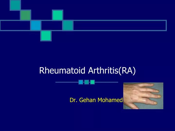
Rheumatoid Arthritis(RA) Dr. Gehan Mohamed
Learning objectives: • At the end of this lecture the student should be able to : • understand definition,genetic predisposition of RA. • Discuss pathophysiology, clinical features of RA. • Identify Diagnostic Criteria ,Laboratory Features and bad prognostic Features of Rheumatoid Arthritis.
RA • Systemic inflammatory autoimmune disorder • Age incidence : 40-70 years of age
Genetics • Patients which have HLA-DRB have Increased risk for : • RA development. • Increased joint damage • Increased joint need for surgery
Macrophages: Produce cytokines Cytokines (TNF-α) cause systemic features Release chemokines recruit PMNs into synovial fluid/membrane TNF-α & IL-1: Proliferation of T cells Activation of B cells Initiates proinflammatory/joint-damaging processes TH-1 cells: Mediate disease processes Activate B cells B cells: Release cytokines Plasma cells that produce Ab Osteoclasts induce: Bone erosion Juxta-articular & Systemic osteoporosis Role of Immunolog in RA
Pathophysiology • Swelling of Synovial lining • Angiogenesis • Pannus formation in form of : • Synovial thickening/hyperplasia • Inflammatory vascularized tissue • Generation of Metalloproteinases • Cytokine release • Infiltration of leukocytes • Change in cell-surface adhesion molecules & cytokines • Destruction of bone & cartilage
Sequence of events : • Proliferation of synovial membrane cells with inflammatory cell infiltrate • Destruction of joints • Disability
1- Diagnostic Criteria • Symmetric peripheral polyarthritis • Morning Stiffness >1 hour • Extraarticular manifestations • Rheumatoid nodules
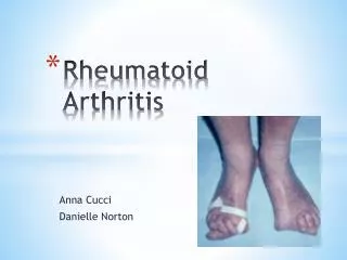
Symmetric Peripheral Polyarthritis • 3 or more Joints for >6 weeks • Intermittent or Migratory involvement • Small Joints • Hands & Feet • Peripheral to Proximal • Leads to Deformity & Destruction of Joints • Erosion of cartilage and bone
Stiffness • Morning or after Prolonged Inactivity • Bilateral • > 1 hours • Reflects severe joint inflammation • Better with movement • Pain with pressure to joint • Pain with movement of joint • Swelling due to hypertrophy of synovium • Effusion • Hottness • Redness
Physical Exam • Decreased grip strength • Carpal tunnel syndrome(condition characterized by pain and numbing or tingling sensations in the hand and caused by compression of a nerve in the carpal tunnel at the wrist. • Ulnar deviation • Boutonniere/Swan neck deformities • Extensor tendon rupture
Myalgia, fatigue, low-grade fever, weight loss, depression. Anemia Rheumatoid nodules Pleuropericarditis Neuropathy Scleritis Splenomegaly Vasculitis Extraarticular Involvement
Rheumatoid Nodules • Extensor surfaces • elbows • Very Specific • Only occur in ~30% • Late in Disease
Arthrocentesis • Confirm diagnoses • Differentiate between inflammatory & noninflammatory • Labs: • White blood cell count if WBC >2000/µL indicates inflammatory arthritis • Gram stain & Culture • Arthroscopy • Evaluate ligamentous & cartilaginous integrity • Biopsy • Infection: aspirate thick
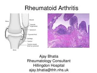
Rheumatoid arthritis : showing inflammatory cell infiltrate in the synovium
Laboratory Features • Rhumatoid Factor • 70-80% of pts. - Lab manifestations up to 10 years before clinical - IgM or IgG - If IgM+ve : more severe disease & poorer outcome. • Overlap with Hepatitis C Virus. • Acute Phase reactants • ESR, CRP monitoring disease activity
Radiology • Evaluate disease activity & joint damage • Bony decalcification
Radiological Studies • Plain Films • Bilateral hands & feet • Color Doppler U/S & MRI • Early signs of damage i.e. Erosions • Bone Edema - even with normal findings on radiography
Mild Disease • Arthralgias • >3 inflamed joints • Mild functional limitation • Minimally elevated ESR & CRP • No erosions/cartilage loss • No extraarticular disease
Moderate Disease • 6-20 Inflamed joints • Moderate functional limitation • Elevated ESR/CRP • Radiographic evidence of inflammation • No extraarticular disease
Severe Disease • >20 persistently inflamed joints • Rapid decline in functional capacity • Radiographic evidence of rapid progession of bony erosions & loss of cartilage • Extraarticular disease
bad prognostic Features • RF +ve • Early development of multiple inflamed joints and joint erosions • Severe functional limitation • Female • HLA epitope presence • Lower socioeconomic status & Less education • Persistent joint inflammation for >12 weeks
Seronegative polyarthritis Psoriatic arthritis Osteoarthritis SLE Paraneoplastic syndrome Crystal-induced arthritis Tophaceous gout Pseudogout Differential diagnosis of arthritis