Download
1 / 13
190 likes | 787 Views
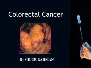
Colorectal Cancer. By GILUR RAHMAN. Introduction. Second most common cause of cancer deaths in the UK. Each year 30,000 new cases diagnosed (68% colon, 32% rectal) Disease more common in westernised countries then Asia or Africa. Aetiology. Age (56% >70yrs old) Colorectal polyps

E N D
Colorectal Cancer By GILUR RAHMAN
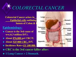
Introduction • Second most common cause of cancer deaths in the UK. • Each year 30,000 new cases diagnosed (68% colon, 32% rectal) • Disease more common in westernised countries then Asia or Africa
Aetiology • Age (56% >70yrs old) • Colorectal polyps • Family history • Genetic • Hereditary non-polyposis colorectal cancer (HNPCC) • Familial adenomatous polyposis (FAP) • Previous colorectal cancer • Ulcerative colitis /colonic crohn’s disease • Diet – hit fat/low fibre • Smoking • Alcohol drinking • Lack of exercise FAP- http://cancerquest.org/images/CancerByType/pics/colon_fap.jpg
Pathology/Pathogenesis • “Adenoma-carcinoma sequence” • Synchronous tumours found in 2% of cases • Most of the tumours found on the left side of the colon • Spread is through local invasion through the bowl wall and via local lymphatics, blood (portal vein into liver) and transcoelomic. • Histology shows well differentiated glandular epithelium with mucin production. Signet rings common characteristics
Symptoms Left sided tumour: • Tenesmus • Blood in stool (fresh red blood) • Obstructive symptoms: • Change in bowl habit • Colicky abdominal pain • Nausea vomiting Right sided tumour: • Weight loss • Anaemia • Abdominal mass (late stage)
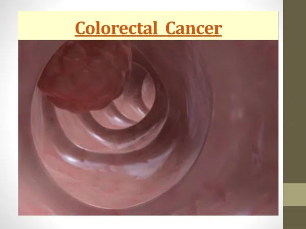
Investigations • Rectal examination • Bloods tests: • - FBC (can show anaemia) • - CEA (carcino embryonic antigen – tumour marker normally used to monitor treatment) • - LFTs (show liver secondaries) • - faecal occult blood • proctoscopy • Barium enema • Sigmoidoscopy – tumours in the last 15cm of GI tract • Colonoscopy – can take biopsy’s • Ultrasound/CT – used to stage look for metastasis (liver) • CT colonography – less invasive then colonoscopies
Staging • Dukes • Type A – tumour confined to mucosa/sub mucose • Type B – invaded through bowl wall but lymph nodes clear • Type C – regional lymph nodes involved • Type D - Distant metastasis
TNM Staging System (Tumor, Node, Metastisis) Tumor • T1: Tumor invades submucosa. T2: Tumor invades muscularis propria. T3: Tumor invades through the muscularis propria into the subserosa, or into the pericolic or perirectal tissues. T4: Tumor directly invades other organs or structures, and/or perforates. Node • N0: No regional lymph node metastasis. N1: Metastasis in 1 to 3 regional lymph nodes. N2: Metastasis in 4 or more regional lymph nodes. Metastasis • M0: No distant metastasis. M1: Distant metastasis present.
Stage Groupings Using the TNM criteria colorectal cancers are placed in to 4 stages: • Stage I: T1 N0 M0; T2 N0 M0 • Stage II: T3 N0 M0; T4 N0 M0 • Stage III: any T, N1-2, M0 • Stage IV: any T, any N, M1 http://homepage.ntlworld.com/watson-jones/portfolio/illustration-08.html
Management Surgery: • Right colon tumours – right hemicolectomy • Transverse colon tumours – extended right hemicolectomy • Descending colon tumours – left hemicolectomy • Sigmoid tumours - Sigmoid colectomy
Right rectal tumour – anterior resection (tumour removed colon anastomosed with remaining rectum) • Low rectal tumour - Abdomioperoneal resection – excise rectum and anus leaving patient with a permanent colostomy
The idea is to remove entire section of bowl supplied by same blood vessel as tumour to ensure clearance of all cancer cells. • Adjuvant chemotherapy increases survival in Dukes B and C. • Neo-adjuvant radiotherapy can be used for rectal tumours, difficult to use with colonic tumours due to the colon not being fixed in a certain position. • Total mesorectal excision (TME) inproves prognosis
Prognosis • Depends upon stage of the cancer • Over 95% survival in dukes A tumours that have been resected • When cancer has spread to lymph nodes far from the colon or rectum, to the lining of the abdominal cavity, or to other organs, the cancer cannot be cured by surgery alone. Survival time is typically only about 7 months