Download
1 / 18
180 likes | 339 Views
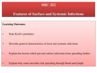
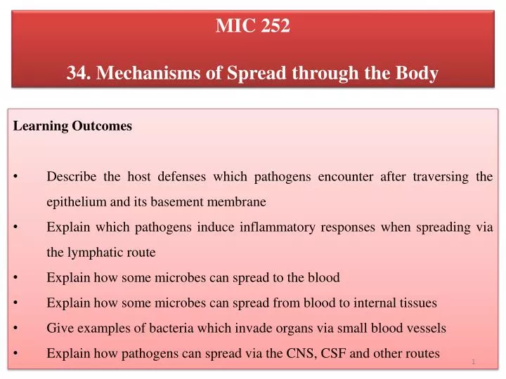
MIC 252 34. Mechanisms of Spread through the Body. Learning Outcomes Describe the host defenses which pathogens encounter after traversing the epithelium and its basement membrane Explain which pathogens induce inflammatory responses when spreading via the lymphatic route

E N D
MIC 252 34. Mechanisms of Spread through the Body • Learning Outcomes • Describe the host defenses which pathogens encounter after traversing the epithelium and its basement membrane • Explain which pathogens induce inflammatory responses when spreading via the lymphatic route • Explain how some microbes can spread to the blood • Explain how some microbes can spread from blood to internal tissues • Give examples of bacteria which invade organs via small blood vessels • Explain how pathogens can spread via the CNS, CSF and other routes
Normal Flora?, Soil, Water, Foul, Rodents, etc Contact, fomites, droplet, vehicle, airborne, arthropods Portals of entry Traverse epithelium, basement membrane, junctions, invade cells, spreading factors Hydrophobicity, surface charge, pili, fimbriae, capsules, LPS, LTA
Invasion of Host Cells and Tissues- Lecture 24 • After adherence, pathogens invade and colonize the host by traversing the epithelium and its basement membrane at the body surface • Some invade tissues through the junctions between epithelial cells • Others invade the cells and so enter the tissue • However, invading microbes face the following defences: • tissue fluids containing antimicrobial substances (antibody, complement); • local macrophages (histiocytes). Subcutaneous and submucosal macrophages are a threat to microbial survival • the physical barrier of local tissue structure. Local tissues consist of various cells in a hydrated gel matrix; although viruses can spread by stepwise invasion of cells, invasion is more difficult for bacteria, and those that spread effectively sometimes possess special spreading factors • the lymphatic system which conveys microorganisms to the battery of phagocytic and immunologic defenses awaiting them in the local lymph node (rare unless following injury or arthropod bite)
Spreading Factors • "Spreading Factors" is a descriptive term for a family of bacterial enzymes that affect the physical properties of tissue matrices and intercellular spaces, thereby promoting the spread of the pathogen. • Hyaluronidase is the original spreading factor produced by streptococci, staphylococci, and clostridia attacks the interstitial cement ("ground substance") of connective tissue by depolymerising hyaluronic acid • Collagenase is produced by Clostridium histolyticum and Clostridium perfringens breaks down collagen, the framework of muscles, which facilitates gas gangrene • Neuraminidase is produced by intestinal pathogens such as Vibrio cholerae and Shigella dysenteriae degrades neuraminic acid (also called sialic acid), an intercellular cement of the epithelial cells of the intestinal mucosa
Spreading Factors • Streptokinase and Staphylokinase are produced by streptococci and staphylococci, respectively converts inactive plasminogen to plasmin digests fibrin prevents clotting of blood relative absence of fibrin in spreading bacterial lesions allows more rapid diffusion of the infectious bacteria • Staphylococcal coagulase cell-associated and diffusible enzyme converts fibrinogen fibrin causes clotting • Coagulase activity is almostalways associated with pathogenic S. aureus and almost never with non-pathogenic S. epidermidis. • I.t.o. virulence, cell bound coagulase could provide an antigenic disguise if it clotted fibrin on the cell surface. • Alternatively, staphylococcal lesion encased in fibrin (e.g. a boil or pimple) could make the bacterial cells resistant to phagocytes or tissue bactericides or even drugs (antibiotics) which might be unable to diffuse to their bacterial target.
Spread in Lymph • Many bacteria that spread along the lymphatic route induce significant signs of inflammation. • E.g., a minor skin wound followed by red streaking and tender swollen local lymph nodes are classical signs of inflamed lymphatic vessels caused by invasion of streptococci • The bacteria secrete toxins or cause other forms of tissue damage that early on in the course of infection set off alarm bells to cause inflammation and alert the non-specific interior defenses • In general, bacterial spread is accompanied by inflammation - bacterial spread is noisy
Minor Wound on Toe with Red Streaking Up the Foot and Ankle Note the minor wound on the toe and red streaking up the foot and ankle. The bacteria are moving through the lymphatic vessels causing inflammation in the vessels.
Spread in Lymph • In contrast to bacteria, many viruses can invade along the lymphaticswithout inducing inflammation, they are silent and asymptomatic • Viruses cause little tissue damage early in the course of infection so inflammatory responses are absent or delayed • E.g., EBV- may be present for weeks before any inflammatory responses are activated • And some viruses can infect cells for long periods of time before they damage them
Spread from Blood • A microbial infection might be arrested at any point during lymphatic spread • But microbes that can evade phagocytes(more detail later) or multiply in the lymph nodes will ultimately reach the blood stream, because movement from one system into the other is easy • Small numbers of bacteria can enter the blood without causing damage • Transient bacteremias, bacteria in the blood, are common • E.g., every time you brush your teeth you get a transient bacteremia • But these bacteria are usually filtered out by the macrophages lining the liver and spleen sinuses
What happens to Bacteria moving through Blood is related to: • Whether they are free in the plasma where they are exposed to phagocytes and antibodies, or • Whether they are intracellular in circulating cells, like erythrocytes, monocytes, lymphocytes, where they can escape immune detection - a more successful strategy • E.g., EBV and Listeria are intracellular in monocytes or lymphocytes, malaria in erythrocytes, Salmonella and Leishmania in macrophages • Microbes can leave the circulation and enter other body sites
Spread from Blood to Internal Tissues • Sometimes free bacteria that enter the blood have the opportunity to colonize internal sites that are less well protected before being eliminated by phagocytes • E.g., such as congenitally abnormal heart valves in the case of viridans streptococci causing infective endocarditis, or in the ends of growing bones in the case of Staphylococcus aureus osteomyelitis • On entering the blood, microorganisms are exposed to macrophages of the reticuloendothelial system • Here in the sinusoids, where blood flows slowly, they are often phagocytosed and destroyed
Spread from Blood to Internal Tissues • But certain microorganisms survive and multiply in these cells (Salmonella typhi, Leishmaniadonovani, yellow fever virus). • The microorganism may then: • spread to adjacent hepatic cells in the liver (hepatitis viruses), or splenic lymphoid tissues (measles virus); • re-invade the blood (S. typhi, hepatitis viruses) • If uptake by reticuloendothelial macrophages is not completewithin a short time, or if large numbers of microorganisms are present in the blood, there is an opportunity for localization elsewhere in the vascular system. • Why each circulating microorganism invades characteristic target organs and tissues is not completely understood, but may be due to:
Spread from Blood to Internal Tissues • specific receptors for the microorganism, leading to localization on the vascular endothelium of certain target organs; • random localization in organs throughout the body, only some of them being suitable for subsequent colonization and replication; • accumulation of circulating microbes in sites where there is local inflammation, because of the slower flow and sticky endothelium in inflamed vessels • After localization and organ invasion, the replicating microbe is shed from the body if the organ has a surface with access to the outside world • It may also be shed back into the bloodstream, either directly or via the lymphatic system
Spread via Nerves • Certain viruses spread via peripheral nerves from peripheral parts of the body to the CNS and vice versa • E.g., Clostridium tetaniTetanus toxinreaches the CNS by this route • Rabies, herpes simplex virus (HSV) and varicella-zoster virus (VZV) travel slowly in axons (10mm/hour), this movement is important in the pathogenesis of these infections • Rabies not only reaches the CNS largely by peripheral nerves, but takes the same route from the CNS when it invades the salivary glands • There are few if any host defenses to control viral spread once the nerves are infected • Viruses and bacteria in the nasopharynx (e.g. meningococci, poliovirus) generally spread to the CNS via the blood • Routes of invasion of the CNS are illustrated in diagram below
Spread via CSF • There is a barrier between the blood and the cerebro spinal fluid and between the blood and brain that is impervious to most microbes (this barrier is called the Blood-Brain Barrier) • But there are some microbes that can cross the blood-brain barrier and gain access to the CSF or brain • These infections are especially dangerous as this barrier also keeps out most host immune cells and drugs • Neisseria meningitidis, Haemophilus influenzae- cause increased permeability of BBB and cross over into the brain
Spread via Other Routes • Both the pleural and peritoneal cavities are lined by macrophages, as if in expectation of such invasion • The peritoneal cavity contains an antimicrobial armory,consisting of the omentum (the 'abdominal policeman'), and many lymphocytes, macrophages and mast cells • Injury or disease in an abdominal organ provides a source of infection for peritonitis, as do chest wounds or lung infections for pleurisy