Download
1 / 1
10 likes | 182 Views
Genetic Correction of Huntington’s Disease Phenotypes in Induced Pluripotent Stem Cells Ningzhe Zhang*, Mahru C. An*, Gary Scott, Daniel Montoro , Tobias Wittkop , Sean Mooney, Simon Melov

E N D
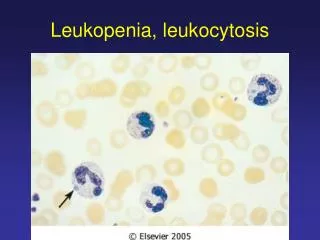
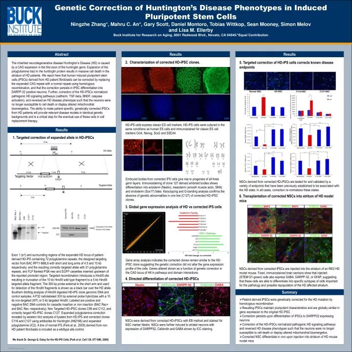
Genetic Correction of Huntington’s Disease Phenotypes in Induced Pluripotent Stem Cells Ningzhe Zhang*, Mahru C. An*, Gary Scott, Daniel Montoro, Tobias Wittkop, Sean Mooney, Simon Melov and Lisa M. EllerbyBuck Institute for Research on Aging, 8001 Redwood Blvd., Novato, CA 94945/*Equal Contribution Abstract Results Results 2. Characterization of corrected HD-iPSC clones. 5. Targeted correction of HD-iPS cells corrects known disease endpoints The inherited neurodegenerative disease Huntington’s Disease (HD) is caused by a CAG expansion in the first exon of the huntingtin gene. Expansion of the polyglutamine tract in the huntingtin protein results in massive cell death in the striatum of HD patients. We report here that human induced pluripotent stem cells (iPSCs) derived from HD patient fibroblasts can be corrected by replacing the expanded CAG repeat with a normal repeat using homologous recombination, and that the correction persists in iPSC differentiation into DARPP-32 positive neurons. Further, correction of the HD-iPSCs normalized pathogenic HD signaling pathways (cadherin, TGF-beta, BNDF, caspase activation), and reversed an HD disease phenotype such that the neurons were no longer susceptible to cell death or display altered mitochondrial bioenergetics. The ability to make patient-specific, genetically corrected iPSCs from HD patients will provide relevant disease models in identical genetic backgrounds and is a critical step for the eventual use of these cells in cell replacement therapy. Nanog Oct4 Sox2 SSEA4 DAPI DAPI DAPI DAPI HD-iPS cells express classic ES cell markers. HD-iPS cells were cultured in the same conditions as human ES cells and immunostained for classic ES cell markers Oct4, Nanog, Sox2 and SSEA4. Results 1. Targeted correction of expanded allele in HD-iPSCs Embryoid bodies from corrected iPS cells give rise to progenies of all three germ layers. Immunostaining of clone 127 derived embrioid bodies shows differentiation into ectoderm (Nestin), mesoderm (smooth muscle actin, SMA) and endoderm (Sox17) fates. Karyotyping and G-banding analysis confirms the absence of genetic abnormalities in one line (C127) of corrected HD-iPSC clones. NSCs derived from corrected HD-iPSCs are tested for and validated by a variety of endpoints that have been previously established to be associated with the HD state. In all cases, correction re-normalizes these states. 6. Transplantation of corrected NSCs into stritum of HD model mice 3. Global gene expression analysis of HD vs corrected iPS cells Exon 1 (e1) and surrounding regions of the expanded HD locus of patient-derived HD-iPS containing 72 polyglutamine repeats; the designed targeting vector from BAC RP11-866L6 with short and long arms of 4.5 and 10 kb respectively; and the resulting correctly targeted allele with 21 polyglutamine repeats, and FLP flanked PGK-neo and EGFP cassettes inserted upstream of the reported promoter region. Targeted recombination introduces a HindIII site resulting in truncation of the 10 kb HindIII wild type fragment to a 5 kb HindIII targeted allele fragment. The 300 bp probe external to the short arm and used for detection of the HindIII fragments is shown as a black bar over the HD allele. Southern blotting analysis of HindIII digested HD-iPS clone genomic DNA and control samples. A P32 radiolabeled 300 bp external probe hybridizes with a 10 kb non-targeted (WT) or 5 kb targeted HindIII. Labeled are positive and negative BAC DNA controls for cassette insertion or non insertion (BAC Rec+ and BAC Rec- respectively); Non Targeted HD-iPSC clones C99 and C112; and correctly targed HD-iPSC clones C127. Expanded polyglutamine correction revealed by western blot analysis of lysates from HD-iPS and corrected clones C116 and C127 using antibodies for Huntingtin (Htt2166) and expanded polyglutamine (IC2). A line of normal iPS (Park et al., 2008) derived from non HD patient fibrobasts is included as a wildtypealle control. Gene array analysis indicates the corrected clones remain similar to the HD-iPSC clone suggesting the genetic correction did not alter the gene expression profile of the cells. Genes altered shown as a function of genetic correction in the CAG locus of Htt in pathways and domain interactions. NSCs derived from corrected iPSCs are injected into the striatum of an R6/2 HD model mouse. Fixed, immunostained brain sections show that injected (STEM121-green) cells also express GABA, DARPP-32, or GFAP, suggesting that these cells are able to differentiate into specific subtypes of cells important for the pathology and possible repopulation of the HD affected striatum. 4. Directed differentiation of corrected HD-iPSCs Summary • Patient derived iPSCs were genetically corrected for the HD mutation by homologous recombination • Resulting iPSCs maintain pluripotent characteristics and are globally similar in gene expression to the original HD iPSC. • Correction persists upon differentiation of iPSCs to DARPP32 expressing neurons • Correction of the HD-iPSCs normalized pathogenic HD signaling pathways and reversed HD disease phenotypes such that the neurons were no longer susceptible to cell death or display altered mitochondrial bioenergetics. • Corrected NSC differentiate in vivo upon injection into striatum of HD mouse model mice NSCs were derived from corrected HD-iPSCs with EB method and stained for NSC marker Nestin. NSCs were further induced to striatal neurons with expression of DARPP32, Calbindin and GABA shown by ICC staining. We thank Dr. George Q. Daley for the HD-iPS Cells (Park et al. Cell 134, 877-886, 2008)