Download
1 / 16
210 likes | 387 Views
The Vascular Cambium. Definitions Cell division related to cambial activity Axial : Along the axis of the organ, or organism Radial : At right angles to the axis, i.e., along a radius Tangential : At right angles to a radius.

E N D
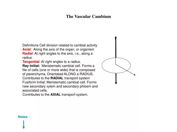
The Vascular Cambium Definitions Cell division related to cambial activity Axial: Along the axis of the organ, or organism Radial: At right angles to the axis, i.e., along a radius Tangential: At right angles to a radius. Ray Initial: Meristematic cambial cell. Forms a file of cells (one or more wide) that is composed of parenchyma. Orientated ALONG a RADIUS. Contributes to the RADIAL transport system Fusiform Initial: Meristematic cambial cell. Forms new secondary xylem and secondary phloem and associated cells. Contributes to the AXIAL transport system. Notes
THE VASCULAR CAMBIUM The vascular cambium is unlike the primary meristems (root and shoot apex) of the plant, in that it produces new cells and tissues which add to the axial system(i.e. the conducting system) as well as to the radial system (i.e. the lateral transport pathway). In contrast, apical meristems of the shoot and root add only to the axial system. The cells of the vascular cambium do not fit the regular concept of meristematic cells (i.e. small, isodiametric shaped cells, with a dense cytoplasm and containing large nuclei). Cambial cells are usually highly vacuolate and occur in two forms, namely fusiform cells and ray cells. Fusiform cells areprism-shaped with a distinct wedge-shape at both ends. Ray cellsareshort and squat. Tangentially, both cell types may be wider than they appear in radial section or longitudinal view. The slides that follow will assist to orientate you with respect to the planes of cell division within the cambium. The two cell types (fusiform and ray cells) have unique functions. Fusiform cells usually only produce cells associated with the axial system -- that is, they produce either new elements of the xylem, or elements of the phloem. Fusiform cells thus add new cells to the AXIAL conducting system. Ray cells on the other hand, produce ONLY ray cells and thus add to the RADIAL system of the plant
AXIAL Axial: Longitudinal translocation, xylem & phloem elements. TANGENTIAL RADIAL Radial: Lateral translocation. Carbohydrate from phloem, to parenchymatic (living) tissue, water from xylem to living tissues as well.
Fusiform vs. ray initials radius Fusiform and ray cells form FILES of cells – each file contains a number of differentiating elements. Both can divide radially OR tangentially tangent endarch TANGENTIAL face exarch TANGENTIAL face exarch TANGENTIAL face RADIAL face RADIAL face plane of cell division normal plane of cell division
During primary growth, the vascular bundles produce PRIMARY vascular tissue. These are the primary phloem (proto + meta) and primary xylem (proto and meta). The fascicular cambium separates the two tissues. Development of secondary vascular tissues in stems Remember: a fascicle is a vascular bundle
Development commences within the fascicular cambium (between the primary phloem and primary xylem) 1.
First activity is in the vascular bundle 2. FCZ = fascicular cambial zone Secondary xylem Secondary phloem FC Secondary xylem and phloem are produced by the fascicular cambium (FC).
3 The interfascicular regions begins to differentiate and a cambium originates here.
PX MX 2X CZ 2P PPF 1P CZ 4 The interfascicular cambial area in herbaceous stems is not usually active, thus does not produce new phloem or xylem tissues 3b 3a A widening band of secondary vascular tissue results from the cambium’s activity.
CZ The ring of secondary tissue is Complete. The interfascicular and fascicular cambia together form a vascular cambium
Phloem Protoxylem Metaxylem endodermis pericycle Typical Dicot Root, end of primary growth (cambial activity in Gymnospermous roots is very similar). Cambial activity in the root
Formation of the cambium. Stage 1. endodermis pericycle Typical Dicot Root cambium appears between the metaxylem and the metaphloem
Formation of the cambium. Stage 2. endodermis pericycle Typical Dicot Root
2X 2P filling out Cambial cells differentiate more rapidly at the interface between the metaxylem and the metaphloem. secondary phloem is produced centrifugally (outwards) as is secondary xylem. In a centripetal (inward) direction. Outward pressure begins to be applied to the primary phloem strands. These strands are gradually forced outwards, to make way for the newly-added secondary vascular tissues.
2X 2P P E 2. Primary phloem strands become crushed, loose functionality 2X 2P P E 3. Primary phloem strands have lost their functionality filling out E = endodermis P = pericycle 2X = secondary xylem 2P = secondary phloem P – pericycle E = endodermis 1. Outward pressure caused by the addition of new cells continues – effectively ‘rounding out’ the ring of secondary vascular tissue
Remnant of primary phloem strands – (located by orange arrows) become completely crushed, and are non-functional E 2P P 2X E = endodermis P = pericycle 2X = secondary xylem 2P = secondary phloem P – pericycle E = endodermis The root will retain its primary xylem, which will be visible and may be functional