Download
1 / 13
950 likes | 2.57k Views
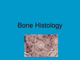
BONE HISTOLOGY. Histology of Bone. Bone consists of widely separated cells surrounded by large amounts of matrix Four principal types of cells: Osteoprogenitor cells – multipotent stem cells that can turn into other bone cells.

E N D
Histology of Bone • Bone consists of widely separated cells surrounded by large amounts of matrix • Four principal types of cells: • Osteoprogenitorcells – multipotent stem cells that can turn into other bone cells. • Osteoblasts – bone forming cells located in peri & endosteum. Synthesize collagen.
Histology of Bone • Osteocytes – mature bone cell (osteoblast surrounded by a calcium matrix). Maintain daily metabolism such as the exchange of nutrients and wastes with the blood. • Osteoclasts – cell that destroys or reabsorbs bone cells and are concentrated in the in the endosteum
Histology of Bone • Matrix contains some calcium carbonate, calcium phosphate but mostly hydroxyapatite, which is the PRIMARY salt that makes bone hard. • Matrix (Bone) composition = 25% water, 25% protein fibers and 50% minerals
What are the tissues that make up bones? 1. Compact Bone Tissue • Outer layer of bone • Dense in appearance • Made out of water, collagen, and crystalized mineral salts (Ca, P) 2. Spongy Bone Tissue • Inner layer of bone • Filled with red bone marrow • Arranged in bars and plates called Trabeculae • Porous openings between trabeculae = spongy appearance
What does compact bone look like at the microscopic level? • Matrix organized into structural units called OSTEONS • aka Haversian System • Circular and tubelike • Arranged into layers (rings) called lamellae • Lamellae are made up of protein fibers, calcium, phosphorus and other minerals • Lamellae surround central canal which contains blood vessels, nerves and lymph
What separates the Lamellae circles? • Rings of osteocytes (mature bone cells) • Osteocytes are found in spaces called lacunae • Tiny canals called canaliculi radiate outward from the central canal to all lacunae making them connected • Blood vessels and nerves enter central canal from periosteum
Bone Growth • Most bone is formed by Endochondral ossification (IN- outward) • Occurs in all bones except flat bones. • Step 1 = Cartilage forms • Step 2 = Cartilage grows • Step 3 = Bone replaces the cartilage from the inside center outward.
Intramembranous • Within membranes, no cartilage precursor • Forms flat bones
What is cartilage? • Special form of dense connective tissue that is comprised of collagen and elastin fibers. • Cartilage is created by chondrocytes. • No blood vessels so nutrients and waste are transported via diffusion. Very slowly.