Download
1 / 37
420 likes | 653 Views
Haematopoiesis and Niche. David Scadden. Gerald and Darlene Jordan Professor of Medicine, Harvard University. co-director . Director . Circuitous career path. 1975 B.A. Bucknell University (English)

E N D
Haematopoiesis and Niche David Scadden
Gerald and Darlene Jordan Professor of Medicine, Harvard University. co-director Director
Circuitous career path • 1975 B.A. Bucknell University (English) • Lab in basement experimenting with chemistry sets, etc. “we basically made bombs” • 1975-1976 Columbia University (Pre-Medical Studies) • 1980 M.D. Case Western Reserve School of Medicine • 2004 A.M. Harvard Medical School (Honorary)
David Scadden’s Lab focuses Niche Haematopoiesis • stem cell number genetic and cell biologic approaches to understand the cell autonomous and extrinsic regulators of stem cell cycling and self-renewal • stem cell localization identified novel mechanisms regulating the ability of the stem cell to engage its proper niche using genetic models and high resolution in vivo imaging • stem cell niche defined key elements of the stem cell niche in the bone marrow and how these features may be modified to improve stem cell number and function.
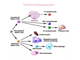
Haematopoiesis • Hematopoiesis -- a process that generates blood cells of all lineages. • Highly regulated process -- to maintain circulating cell numbers within relatively constant levels and to respond rapidly to conditions requiring extra cells . • Normal hematopoiesis involves an highly regulated balance between self-renewal, terminal differentiation, migration, and cell death.
The Hematopoietic stem cell • HSCs are pluripotent • Capable of self-renewal :- production of additional HSCs • Differentiation :- hematopoietic lineages • HSCs are rare (0.01 % of nucleated cells in adult bone marrow) • HSCs are mainly quiescent cells • reside in specialized regulatory microenvironment or • niche where they receive appropriate support for maintaining self renewal and multi-lineage differentiation capacity Difficulty in isolation and maintenance of HSCs a major hurdle
Lkb1 a tumor suppressor Encodes Serine/threonine kinase that links metabolism and cell growth Lkb1 AMPK key regulatory proteins (metabolism) restore cellular ATP levels during energy stress • regulates energy homeostasis and cell structure
Lkb1 deletion causes bone marrow failure Mx1-cre strain Lkb L/L X Mx1-cre; Lkb1L/L mice (designated Lkb1 mutants) On induction with pIpC • progressive pancytopenia • rapid loss of bone marrow myeloid, B lymphoid and erythroid cells
Lkb1 function in bone marrow is cell intrinsic Mx1-cre; Lkb1L/L Wild type Bone marrow transplant pIpC treatment
Lkb1 maintains HSC quiescence Mx1-cre; Lkb1L/L Bone marrow at day -3 increase in HSCs Bone marrow at day +2 marked reduction pIpC Cell cycle analysis using Ki-67 and propidium iodide after pIpC induction • Lkb1 is required for the maintenance of HSC quiescence and its loss causes bone marrow failure that is preceded by an increase in the proliferation and absolute number of HSCs.
Lkb1 loss induces apoptosis and autophagy (LC3 levels) Viability (7AAD staining) Cleaved caspase 3 levels Lkb1 maintains bone marrow energy homeostasis Mitochondrial membrane potential in the Lkb1 CMPs, (CLP) and HSCs at day 3 after pIpC treatment basal mitochondrial oxygen consumption and total mitochondrial oxidative capacity
CONCLUSION • Lkb1 is needed to maintain haematopoietic cells • broadly, with Lkb1 deletion leading to the death of multiple subpopulations. • processes involved metabolic homeostasis • associated with mitochondrial dysfunction, ATP depletion, and rapid apoptotic cell death. • Lkb1 function integrating energy sensing and growth control • Its unique role in enforcing quiescence of HSCs raises interesting issues of how this population may be particularly responsive to its metabolic environment. Whether this is due to a primary low-energy state in HSCs that activates Lkb1 signalling or the distinct regulation of Lkb1 in these cells is a subject of further study.
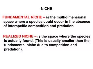
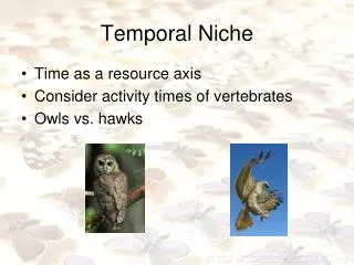
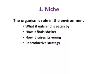
Definition of niche Architecture :- A recessed space ( decorative) Ecology:- A habitat where an organism resides, thrives and reproduce. More formally, the niche includes how a population responds to the abundance of its resources and enemies (functional)
NICHE • Stable environment for stem cells • Niches are subsets of tissues and extracellular subsets that can indefinitely • house stem cells and control their self-renewal and progeny production in vivo
Hematopoietic Stem Cell Niche HSC function is tightly controlled to maintain hematopoietic homeostasis, and this regulation relies on specialized cells and factors that constitute the hematopoietic ‘niche’, or microenvironment Components of a hypothetical HSC ‘niche’. Leo D. Wang and Amy J. Wagers, Nature Reviews Molecular Cell Biology ,2011
Concept of niche in malignant transformation • Pioneering insights into the cellular hierarchy of blood cell formation, and the unique ability to interrogate well defined subsets of hematopoietic cells have lead to the identifying molecular events leading to its malignant transformation. • The reductionist, hematopoietic cell-centered approach to the understanding of hematopoiesis and its deregulation in hematopoietic cancer has its limitations. • The early events driving the initiation and malignant evolution of pre-malignant states remain largely unknown.
MicroRNA BIOGENESIS RNA-induced silencing complex (RISC) Kim et al.,Nature Rev.6 ,376-385(2005)
Mouse model • Osx-GFP-Cre+ Dicer fl/+ (OCDfl/+ control) and • Osx-GFP-Cre+ Dicerfl/fl (OCDfl/fl mutant )mice. • crossing mice expressing green fluorescent protein (GFP)-Crerecombinase driven by the Osterixpromoter (expressed first in osteoprogenitor cells) with mice with floxed Dicer1 alleles(OCDfl/fl mice). • Mutant mice were showed growth retardation and impaired survival (30% mutant mortality by 8 weeks). Therefore, examined mice at 4–6 weeks of age.
Impaired osteoblastic differentiation(In vivo) E.OCDfl/fl bone marrow stromal cells -reduced CFU-ALK F, osteoblasts from OCDfl/fl mice expressed less of the terminal differentiation marker osteocalcin Arrows in H indicate osteocalcin at the endosteal surface
Myelodysplasia in OCDfl/fl mice B.Blood smears showing dysplastic hyperlobulated nuclei in granulocytes C.Bone marrow sections showing micro-megakaryocytes with hyperchromatic nuclei A.Leukopenia,thrombocytopenia Peripheral cytopenia with dysgranulopoiesis and dysplastic megakaryocytes is consistent with a diagnosis of myelodysplastic syndrome (MDS) in mice according to the Bethesda criteria.
Myelodysplasia is environmentally induced Transplantation to assess the contribution of the microenvironment to the haematopoietic phenotype Cytopenia Complete normalization of leukopenia (c) granulocyte and (d)megakaryocyte morphology
Myelodysplasia is environmentally induced (f)Haematopoiesis in mutant mice at 8 weeks showed leukopenia (g)dysgranulopoiesis with giant platelets ( indicated by arrows) (h)increased bone marrow vascularity with dysplastic megakaryopoiesis( arrows).
Deletion of Shwachman–Diamond–Bodian syndrome gene • To obtain insight into the genes and molecular pathways in osteolineage cells driving the disruption of haematopoiesis in OCDfl/fl mice- performed gene expression profiling of primary OCDfl/+ and OCDfl/fl osteolineage cells • significant downregulation of the Shwachman–Diamond–Bodian syndrome (Sbds) gene a.Genetic model of Sbds deletion from osteoprogenitor cells b, Leukopenia c.Dysplasiaof neutrophils in peripheral blood. d. Dysplasia of megakaryocytes (micromegakaryocytes). e. Hypervascularity of the bone marrow. f. Increased intramedullary apoptosis of haematopoietic progenitor cells
Dysfunctional Niches as a Root of Hematopoietic Malignancy Shiozawa et al.,Cell Stem Cell 6, May 7, 2010
Autologous Stem cell Transplantation Thalassaemia, Leukemia, Multiple Sclerosis, etc.,
Groups in HSPC Mobilization HSC Mobilization Good Mobilizers Poor Mobilizers ? Reason --- Unknown
Mice Models • Type I – Streptozotocin • Type II – (db/db) LEPR knockout.