Download
1 / 29
290 likes | 458 Views
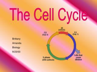
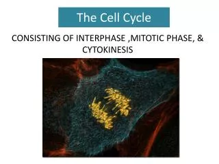
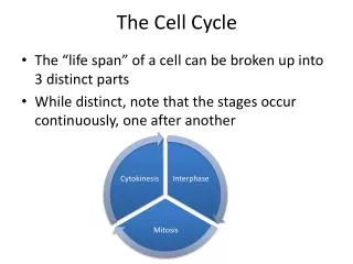
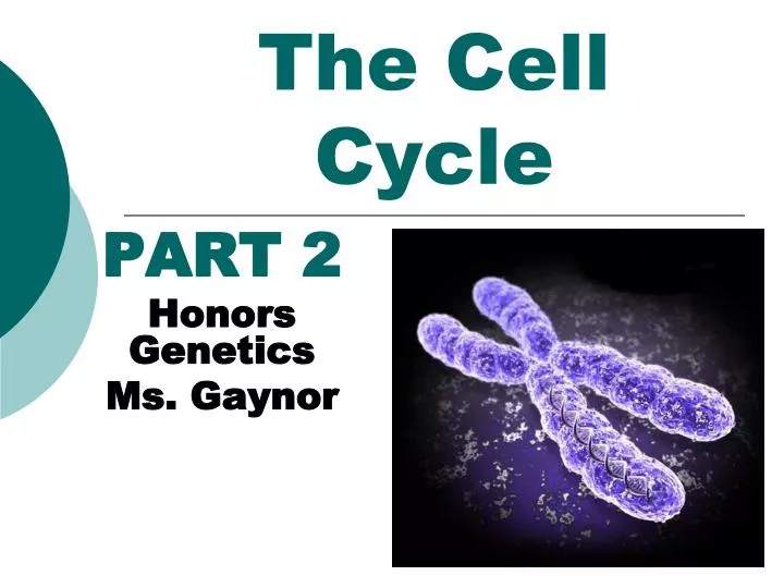
The Cell Cycle. PART 2 Honors Genetics Ms. Gaynor. The Cell Cycle. The mitotic phase alternates with interphase in the cell cycle Interphase mitosisinterphasemitosis. INTERPHASE. S (DNA synthesis). G 1. Cytokinesis Mitosis. G 2. MITOTIC (M) PHASE. Figure 12.5.

E N D
The Cell Cycle PART 2 Honors Genetics Ms. Gaynor
The Cell Cycle • The mitotic phase alternates with interphase in the cell cycle • Interphasemitosisinterphasemitosis
INTERPHASE S(DNA synthesis) G1 CytokinesisMitosis G2 MITOTIC(M) PHASE Figure 12.5 Phases of the Cell Cycle • The cell cycle consists of • The mitotic (M) phase • Interphase (90% of the cell’s life)
Interphase can be divided into subphases • G1 phase (GAP 1 phase) • cell grows in size • varies most in length from cell to cell • S phase (synthesis phase) • DNA is copied (DNA replication) • Single Double • Other organelles are copied (ex: centrosomes in animal cells) • G2 phase (GAP 2 phase) • More growth and preparation (make proteins) for mitosis http://www.cellsalive.com/cell_cycle.htm
Another G phase of Interphase • Called G0 phase called the resting phase • The cell exits the “cycle” and (usually) does NOT reproduce again • Ex: muscle cells, nerve cells, red blood cells
Interphase“Intermission” or “Inbetween” • not part of mitosis • Includes stages G1, S, and G2 of the cell cycle • DNA is in chromatin form • Nucleus & nucleolus present • Longest phase of cell cycle
The Mitotic (M) phase • Is made up of 2 parts 1. Mitosis division of the nucleus (called Karyokinesis) 2. Cytokinesis division of the cytoplasm
Mitosis • Continuous pathway (Early, Mid, & Late) • Consists of 4 phases and cytokinesis • Prophase • Metaphase • Anaphase • Telophase • Cytokinesis
Prophase (X’s)“Pack Together” • Chromatin Chromosomes • DNA “packs” together • Mitotic spindle fibers form from centrosomes • Centrioles are in centromsomes in animals • appear as asters in animals • Microtubule Organizing Centers (MTOC’s) (in plants) • Centrosomes & Spindle fibers move towards “poles” • Late:Nucleus and nucleolus disappear • kinetochore fibersattach to each kinetochore on each chromosome they begin to migrate toward the cell center
PROMETAPHASE G2 OF INTERPHASE PROPHASE Aster Centrosomes(with centriole pairs) Fragmentsof nuclearenvelope Kinetochore Early mitoticspindle Chromatin(duplicated) Centromere Nonkinetochoremicrotubules Kinetochore microtubule Nucleolus Chromosome, consistingof two sister chromatids Figure 12.6 Nuclearenvelope Plasmamembrane
Mitotic Spindle Fibers • Two types of spindle fibers • Kinetochore fibers • Polar fibers
Metaphase (X’s)“Meet in the Middle” • Chromosomes line up in the middle of cell • Called equatorial or metaphase plate • Kinetochore spinder fibers pull and tug chromosomes to line up
Anaphase (V’s)“Adios and Away” • SISTER CHROMATIDS separate and begin moving to opposite ends (poles) of the cell • Polar fibers lengthen and elongate the cell • Late anaphase each pole contains a complete set of single chromosomes • NOTE: This is the only time there is DOUBLE the amount of DNA in ONE cell
1 Kinetochore Spindlepole Figure 12.8 • In anaphase, sister chromatids separate • And move along the kinetochore microtubules toward opposite ends of the cell
METAPHASE ANAPHASE TELOPHASE AND CYTOKINESIS Metaphaseplate Cleavagefurrow Nucleolusforming Nuclear envelopeforming Daughter chromosomes Centrosome at one spindle pole Spindle Figure 12.6
Telophase (V’s)“Two New Cells” • The spindle fibers dissappear • Two daughter nuclei and nucleoli begin to form at the two poles • Nuclear envelopes reforms from the fragments of the parent cell’s nuclear envelope and other portions of the endomembrane system • Chromosomes chromatin
Cytokinesis“Division of the Cytoplasm” • Occurs in Late telophase • In animal cells • a cleavage furrow forms, which pinches the cell in two. • In plant cells • vesicles from the Golgi apparatus produce a cell plate at the middle of the cell • At the end of cytokinesis, there are two distinct IDENTICAL daughter cells.
Cleavage furrow 100 µm Contractile ring of microfilaments Daughter cells (a) Cleavage of an animal cell (SEM) Figure 12.9 A Cytokinesis: A Closer Look • In animal cells • Cytokinesis occurs by a process known as cleavage, forming a cleavage furrow
Vesiclesforming cell plate Wall of patent cell 1 µm Cell plate New cell wall Daughter cells Figure 12.9 B (b) Cell plate formation in a plant cell (SEM) • In plant cells, during cytokinesis • A cell plate forms
PROMETAPHASE G2 OF INTERPHASE PROPHASE Aster Centrosomes(with centriole pairs) Fragmentsof nuclearenvelope Kinetochore Early mitoticspindle Chromatin(duplicated) Centromere Nonkinetochoremicrotubules Kinetochore microtubule Nucleolus Chromosome, consistingof two sister chromatids Figure 12.6 Nuclearenvelope Plasmamembrane
METAPHASE ANAPHASE TELOPHASE AND CYTOKINESIS Metaphaseplate Cleavagefurrow Nucleolusforming Nuclear envelopeforming Daughter chromosomes Centrosome at one spindle pole Spindle Figure 12.6
2 3 5 1 4 Chromatinecondensing Nucleus Chromosome Nucleolus Metaphase. The spindle is complete,and the chromosomes,attached to microtubulesat their kinetochores, are all at the metaphase plate. Prophase. The chromatinis condensing. The nucleolus is beginning to disappear.Although not yet visible in the micrograph, the mitotic spindle is staring to from. Prometaphase.We now see discretechromosomes; each consists of two identical sister chromatids. Laterin prometaphase, the nuclear envelop will fragment. Telophase. Daughternuclei are forming. Meanwhile, cytokinesishas started: The cellplate, which will divided the cytoplasm in two, is growing toward the perimeterof the parent cell. Anaphase. Thechromatids of each chromosome have separated, and the daughter chromosomesare moving to the ends of cell as their kinetochoremicrotubles shorten. Figure 12.10 Mitosis in a plant cell
Cell Cycle and Mitosis Animations • http://www.ucopenaccess.org/courses/APBiologyI/course%20files/multimedia/lesson17/lessonp.html • http://highered.mcgraw-hill.com/olcweb/cgi/pluginpop.cgi?it=swf::535::535::/sites/dl/free/0072437316/120073/bio14.swf::Mitosis%20and%20Cytokinesis • http://www.sumanasinc.com/webcontent/animations/content/mitosis.html • http://www.johnkyrk.com/mitosis.html
Remember… • “IPMATc” • I peed on the mat, see. • Let’s do the Mitosis Hand Cheer!