Download
1 / 8

E N D
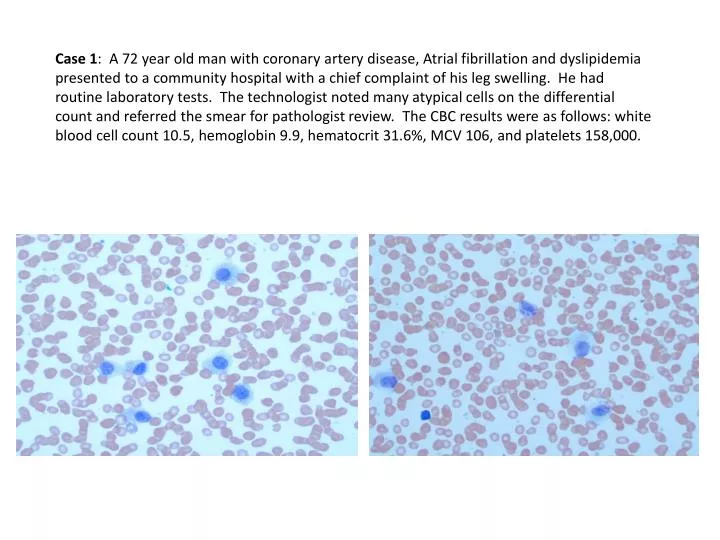
Case 1: A 72 year old man with coronary artery disease, Atrial fibrillation and dyslipidemia presented to a community hospital with a chief complaint of his leg swelling. He had routine laboratory tests. The technologist noted many atypical cells on the differential count and referred the smear for pathologist review. The CBC results were as follows: white blood cell count 10.5, hemoglobin 9.9, hematocrit 31.6%, MCV 106, and platelets 158,000.
Case 2: 67-year-old male with two left renal masses in a pelvic kidney found incidentally on an abdominal CT done for abdominal pain. The patient complained of chronic non-specific abdominal pain for the past several months associated with slight weight loss with no change in appetite. He has been on immune-modulating therapy for Rheumatoid Arthritis for the past 6 months and is a current everyday smoker -- 1.0 packs/day for 50 years. Gross: Partial nephrectomy weighing 45 grams and measuring 5 x 4 x 3.5 cm. Two separate renal masses simulating “camel humps” , one measures 3 x 3 x 2.5 cm, the second 3 x 2.5 x 2.5 cm Mass A Mass B
Case #2 Mass A
Mass B Case #2
Case 3: 49 year old Chinese speaking female presents to ENT clinic with right ear pain, tinnitus and decreased hearing for the last 6 months. More recently she has been experiencing nasal congestion R>L, throat pain and an enlarging right neck mass. On exam:Patient has raspy voiceRight ear: swelling below and in front of right ear, canal is narrow, inflamed and dry with no drainage; tympanic membrane- inflamed bulging irregularity, no perforationNose: Mucosa moist, inflamed with enlarged turbinates R>L; Neck: Supple, 5 x 6 cm mass right neck; Nasopharyngoscopy: exophytic mass in right nasopharynx with erythema FNA PAP FNA diff quick Bx H&E’s
Case 4: 61 yo female presents with history of hypothyroidism for 5 years and suspicious thyroid nodule in the left lobe. History is significant for cerebellar hemangioblastoma s/p resection in August 2013. Her thyroid function test revealed her to be euthyroid with a TSH of 1.3 and a free T3 of 2.8; on 50 mcg of levothyroxine daily. Repeated thyroid FNAs were non-diagnostic, so she underwent total thyroidectomy which showed a well-defined ovoid soft lesion in the mid portion of the left lobe measuring 3.4 cm
Case 5: 70 year old male with past history of anal condylomas, testicular seminoma treated with surgery and radiotherapy, and urothelial carcinoma treated with cystoprostatectomy and chemotherapy. He presented with anal ulcer of two month duration associated with mild intermittent rectal bleeding. A biopsy showed squamous cell carcinoma for what he underwent an abdomino-perineal resection Anal ulcer, resection specimen, 20x Anal ulcer, resection specimen, 20x p16 BCL-2 BerEP4 SOX-2
Case 6: 70 yomale smoker 20 cigarettes / day for more than 30 years presents with hemoptysis. The patient had a past medical history of bilateral femoral-popliteal bypass. Physical exam demonstrates right knee edematous, with inability to flex. CT: knee synovitis suggests organized collection and haemarthrosis. Patient has fever, 38.5Cº. Labs reveal Hb 6.8gr/dl TTF-1 CK