Download
1 / 35
420 likes | 1.15k Views
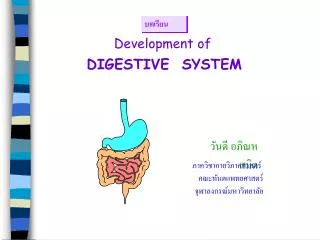
Development of DIGESTIVE SYSTEM. วันดี อภิณหสมิต. ภาควิชากายวิภาคศาสตร์ คณะทันตแพทยศาสตร์ จุฬาลงกรณ์มหาวิทยาลัย. บทเรียน. 1. Oral cavity, tonsils, salivary glands, Pharynx, 2. Esophagus 3. Stomach 4. Duodenum(proximal to bile duct opening) 5. Liver & biliary apparatus 6. Pancreas

E N D
Development of DIGESTIVE SYSTEM วันดี อภิณหสมิต ภาควิชากายวิภาคศาสตร์ คณะทันตแพทยศาสตร์ จุฬาลงกรณ์มหาวิทยาลัย บทเรียน
1.Oral cavity, tonsils, salivary glands, Pharynx, 2. Esophagus 3. Stomach 4. Duodenum(proximal to bile duct opening) 5. Liver & biliary apparatus 6. Pancreas upper respiratory system Lower respiratory system Spleen Foregut Midgut 1. Small intestine 2. Caecum 3. Vermiform appendix 4. Ascending colon, Right 1/2-2/3 transverse colon 1. Right 1/2-12/3 transverse colon 2. Decending colon, sigmoid, rectum, anal canal 3. Epithelium of urinary bladder, urethra Hindgut (Grant’s Atlas of Anatomy, 7th ed.)
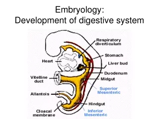
Yolk sac Yolk sac Presomite 22 d • การพัฒนาเริ่มในสัปดาห์ที่ 4 • Embryo ยกตัวสูงขึ้น เกิดhead และ tail folds • Endodermที่บุ yolk sac incorporate เข้าไปใน embryo • เกิดเป็น primordial (primitive, endodermal) gut tube DIGESTIVE SYSTEM (Langman’s Medical Embryology, 8th ed.)
24 d 1 mo. • Primitive gut tube เจริญไปเป็น epithelium และ glands • Epithelium ที่ cranial extremity ของ tractstomodeum (primitive mouth) • Epithelium ที่ caudal extremity ของ tract proctodeum(anal pit) • Muscle, connective tissueและชั้นอื่น ๆ พัฒนามาจาก splanchnic mesoderm (Langman’s Medical Embryology, 8th ed.)
4-wks (Moore & Persaud: The Developing Human: Clinically Oriented Embryology, 6th ed.)
Primordium of liver (hepatic diverticulum) Primitive gut tube 1. Foregut จาก oral membrane ถึงบริเวณที่พบ hepatic diverticulumซึ่งตรงกับ anterior intestinal portal 2. Midgutจาก anterior intestinal portal ถึง posterior intestinal portal ต่อกับ yolk sac 3. Hind gutจากanterior intestinal portal ถึง cloacal membrane duct (Moore & Persaud: The Developing Human: Clinical Oriented Embryology, 6th ed.)
Celiac a. Inferior mesenteric a. Superior mesenteric a. (Netter, Atlas of Human Anatomy)
Celiac a. Inf. mesenteric a. Sup. mesenteric a. (Netter, Atlas of Human Anatomy)
(Tracheoesophageal septum) 3 wks 4 wks Esophagus & Respiratory diverticulum Esophagus เจริญมาจาก foregut ส่วนที่อยู่ caudal ต่อ primitive pharynx 5 wks 4 wks (Langmann’s Medical Embryology, 8th ed.)
Esophagus • เริ่มแรก esophagus จะสั้น • ต่อมาจะยาวออกอย่างรวดเร็ว เนื่องจากการเจริญและเคลื่อนตัวลงต่ำของ • heart & lung • 7 weeks : epithelium เพิ่มจำนวน ท่อตัน • ปลาย 8 weeksเกิด recanalization • Muscularis externa • Superior 2/3เป็น striated muscle มาจาก mesenchyme ใน • caudal pharyngeal arches • Inferior 1/3 เป็น smooth m. มาจาก splanchnic mesenchme
Esophageal atresia ระงับการสร้าง esophagus deviation of tracheopharyngeal septum ไปทางด้านหลัง Esophageal stenosis lumen แคบ เกิดได้หลายแห่ง มักพบที่ distal 1/3 มักเกิดจาก incomplete canalization Short esophagus ไม่มีการยืดยาวออก ทำให้ stomach อยู่ใน thorax
28 d กลาง 4 wks ส่วนปลายของ foregut เริ่มขยายตัวเป็นรูปกระสวย อยู่ในแนวกลาง 35 d ขยายตัวในแนวหน้า-หลัง Stomach (Moore & Persaud: The Developing Human: Clinical Oriented Embryology, 6th ed.)
6 wks ขอบทาง dorsal เจริญเร็วเป็น greater curvature 40 d ในขณะที่ stomach ขยาย เกิด rotation อย่างช้า ๆ รอบแนวแกน ทำมุม 90 องศา 48 d (Moore & Persaud: The Developing Human: Clinical Oriented Embryology, 6th ed.)
ขยายตัว หมุนรอบแนวแกน 90 องศา Rotation of the stomach 1. ขอบทางด้าน ventral (lesser curvature) ด้านขวา ขอบทางด้าน dorsal (greater curvature) ด้านซ้าย 2. ด้านซ้าย ด้าน ventral ด้านขวา ด้าน dorsal 3. ก่อนการหมุน cranial & caudal ends ของ stomach อยู่ในแนวกลาง ระหว่างการหมุน cranial ends เคลื่อนไปทางซ้าย และลงล่าง caudal ends เคลื่อนไปทางขวา และขึ้นบน หลังการหมุนแนวแกนของ stomach ทอดอยู่ในแนวขวางลำตัว 4. การเจริญและการหมุนตัวของ stomach ช่วยอธิบายว่าทำไมในผู้ใหญ่ vagus nerve ข้างซ้ายจึงเลี้ยงผนังทางด้านหน้าของ stomach vagus nerve ข้างขวาจึงเลี้ยงผนังทางด้านหลังของ stomach (Moore & Persaud: The Developing Human: Clinical Oriented Embryology, 6th ed.)
Mesenteries of the stomach 1. Dorsal mesogastrium (Dorsal mesentery) 2. Ventral mesogastrium(Ventral mesentery) (Moore & Persaud: The Developing Human: Clinical Oriented Embryology, 6th ed.)
Omental bursa (lesser peritoneal sac) • Clefts in dorsal mesogastrium • Coalescence ของ clefts เกิดเป็น • omental bursa 5 wks • Dorsal mesentery ขยายตัว • Stomach ขยายตัว • Omental bursa มีขนาดใหญ่ขึ้น • Omental (epiploic) foramen • Inferior recess of omental bursa • ยาวขึ้น เรียกว่าGreater omentum • ต่อมา recess หายไป • เนื่องจากการรวมตัวของชั้นของ • greater omentum H6 wks (Moore & Persaud: The Developing Human: Clinical Oriented Embryology, 6th ed.)
5 wks 4 wks Duodenum • 4 weeks: • Duodenum พัฒนาจาก • 1. Caudal part of foregut • 2. Cranial part of midgut • 3. Splanchnic mesenchyme 5 wks • เจริญเร็วเกิดเป็น loop รูปตัว C • ทอดมาทางด้าน ventral • เมื่อ stomach หมุน • duodenual loop หมุนไปทาง • ขวาไปอยู่ retroperitoneum 5 & 6 weeks: lumen ของ duodenum แคบ และอุดตันชั่วคราว 5 wks 6 wks (Moore & Persaud: The Developing Human: Clinical Oriented Embryology, 6th ed.)
Duodenal stenosis Duodenal atresia Normal lumen 8 weeks: Vacuolation เกิดท่อ (Moore & Persaud: The Developing Human: Clinical Oriented Embryology, 6th ed.)
C Q Liver 1. Right lobe 2. Left lobe 3. Caudate lobe 4. Quadrate lobe (Netter: Atlas of Human Anatomy)
4 wks 5 wks Liver, gall bladder, biliary duct system 4 weeks Hepatic diverticulum อยู่หน้าต่อ caudal part ของ foregut : ยื่นเข้าไปใน septum transversum ซึ่งเป็นส่วนของ splanchnic mesoderm และจะเจริญไปเป็น central tendon of diaphragm ventral mesentery Moore & Persaud: The Developing Human,Clinically Oriented Embryology, 6th ed)
5 wks 4 wks • Hepatic diverticulum • Larger cranial part เป็น • primordium ของliver • Small caudal part เป็น • gall bladder • Stalk of diverticulum เป็น • cystic ducts 5-10 weeks • Liver เจริญเร็วมาก จนเต็ม abdomen • เริ่มแรก Rt และ Lt lobe มีขนาดใกล้เคียงกัน • ต่อมา Rt lobe ยนาดใหญ่กว่า Lt lobe • Caudate & quadrate lobe เจริญมาจาก • Rt lobe • 6 weeks มี Hematopoiesis • 12 weeks เริ่มสร้าง bile 6 wks 5 wks (Moore & Persaud: The Developing Human: Clinical Oriented Embryology, 6th ed.)
5 wks Ventral mesetery 1. Lesser omentum จาก liver ไป lesser curvature of stomach 2. Falciform ligament จาก liver ไป anterior abdominal wall พบ umbilical vein ทอดใน free border ของ falciform ligament ventral mesentery ประกอบเป็น visceral peritoneum ของ liver คลุมทุกส่วนของ liver ยกเว้น bare area (Moore & Persaud: The Developing Human: Clinical Oriented Embryology, 6th ed.)
Pancreas (Netter: tlas of Human Anatomy)
Pancreas: 5-8 weeks • Dorsal pancreatic bud • Ventral pancreatic bud • เมื่อ duodenum หมุน VPB • เคลื่อนไปอยู่ dorsal ต่อ bile duct • VPB รวมกับ DPB (Moore & Persaud: The Developing Human: Clinical Oriented Embryology, 6th ed.)
Spleen • Large vascular lymphatic organ • 5 weeks • เจริญมาจาก mesenchymal cells • ที่อยู่ระหว่าง dorsal mesogastrium • ขณะที่ stomach หมุนตัว • ผิวทางด้านซ้ายของ mesogastrium • รวมกับ peritoneum เหนือ Lt kidney (Moore & Persaud: The Developing Human: Clinical Oriented Embryology, 6th ed.)
MIDGUT 1. Small intestine 2. Caecum 3. Vermiform appendix 4. Ascending colon, Right 1/2-2/3 transverse colon
10 weeks Rotation of midgut • Midgut ยืดยาวออก • เป็น midgut loop ี่ • ที่เป็นรูปตัว U • Midgut loop ยื่นเข้าไป • ใน extraembryonic • coelom ของ umbilical cord • เรียกว่า physiological • umbilical herneation • (6wks) • Loop ยึดกับ post. • abdominal wall ด้วย • elongated mesentery 6 weeks • Midgut loop • 1. Cranial limb small intestinal loop • 2. Caudal limb ไม่ค่อยเปลี่ยน • caecal diverticulum • Midgut loop หมุนทวนเข็มนาฬิกา 90 องศา • รอบ superior mesenteric artery • Cranial limb ไปอยู่ด้านขวา • Caudal limb ไปอยู่ด้านซ้าย (Moore & Persaud: The Developing Human: Clinical Oriented Embryology, 6th ed.)
11 weeks Later stage Returning of the midgut to abdomen 10 wks - liver & kidneys ขนาดเล็กลง - Abdominal vavity ขยาย Reduction of physiological midgut hernia Small intestine -กลับเข้าไปก่อน และอยู่หลังต่อ sup mesenteric a Large intestine -หมุนทวนเข็มนาฬิกา 180 องศา (Moore & Persaud: The Developing Human: Clinical Oriented Embryology, 6th ed.)
Fixation of intestines ภายหลังการหมุนของ stomach & duodenum Duodenum (midgut) & pancreas กดบน posterior abdominal wall เกิด fusion retroperitoneal position Before 4 mo 1. ภายหลังการหมุนของ intestine Ascending & descending colon กดบน posterior abdominal wall เกิด fusion retroperitoneal position After 2. Mesentery ของ transverse colon รวมกับ posterior wall ของ greater omentum 3.Jejunum และ Ileum ยังคงมี mesentery ย้ายจากที่เกาะกับ ascending colon มา เกาะที่ duodenum (intraperitoneum) ไปยัง ileocecal junction New born (Moore & Persaud: The Developing Human: Clinical Oriented Embryology, 6th ed.)
8 wks 12 wks 6 wks Adult At birth • หลังคลอด ผนังของ cecum เจริญไม่เท่ากัน • Appendix เคลื่อนไปอยู่ด้าน medial • Ascending colon ยาวขึ้น • appendix ไปอยู่ retrocecal Caecum and Vermiform appendix (Moore & Persaud: The Developing Human: Clinical Oriented Embryology, 6th ed.)
Umbilical cord Site of liver in sac Intestine Anterior abdominal wall Persistence of the herniation of abdominal contents in the proximal portion of umbilical cord (Moore & Persaud: The Developing Human: Clinical Oriented Embryology, 6th ed.)
HINDGUT 1. Right 1/2-12/3 transverse colon 2. Decending colon, sigmoid, rectum, anal canal 3. Epithelium of urinary bladder, urethra
Cloaca • ส่วนปลายของ hindgut • บุด้วย endodermที่รวม • กับ surface ectodermที่ • cloacal membrane 4wks • urorectal septum • (mesenchyme ที่เจริญอยู่ระหว่าง • allantois และ hindgut) • แบ่งเป็น dorsal และ ventral • parts • 1. Dorsal :rectum & cranial • part of anal canal เจริญไป • เป็น urinary bladder & urethra • 2.Ventralเป็น urogenital sinus • เจริญไปเป็น rectum & sup. • part of anal canal 6 wks 8 weeks Anal membraneขาด 7 wks (Moore & Persaud: The Developing Human: Clinical Oriented Embryology, 6th ed.)
Anal Canal • Superior 2/3 เจริญมาจาก hindgut • Inferior 1/3 เจริญมาจาก proctodeum