Download
1 / 97
980 likes | 989 Views
Fundamentals of the Nervous System and Nervous Tissue: Part A. *Functions of the Nervous System. Sensory input Information gathered by sensory receptors about internal and external changes Integration Interpretation of sensory input Motor output

E N D
Fundamentals of the Nervous System and Nervous Tissue: Part A
*Functions of the Nervous System • Sensory input • Information gathered by sensory receptors about internal and external changes • Integration • Interpretation of sensory input • Motor output • Activation of effector organs (muscles and glands) produces a response
Sensory input Integration Motor output Figure 11.1
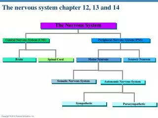
*Divisions of the Nervous System • Central nervous system (CNS) • Brain and spinal cord • Integration and command center • Peripheral nervous system (PNS) • Paired spinal and cranial nerves carry messages to and from the CNS
Peripheral Nervous System (PNS) • Two functional divisions • Sensory (afferent) division • Somatic afferent fibers—convey impulses from skin, skeletal muscles, and joints • Visceral afferent fibers—convey impulses from visceral organs
* • Motor (efferent) division • Transmits impulses from the CNS to effector organs
*Motor Division of PNS • Somatic (voluntary) nervous system • Conscious control of skeletal muscles
Peripheral nervous system (PNS) Central nervous system (CNS) Cranial nerves and spinal nerves Brain and spinal cord Communication lines between the CNS and the rest of the body Integrative and control centers Sensory (afferent) division Motor (efferent) division Somatic and visceral sensory nerve fibers Motor nerve fibers Conducts impulses from the CNS to effectors (muscles and glands) Conducts impulses from receptors to the CNS Somatic sensory fiber Autonomic nervous system (ANS) Somatic nervous system Skin Visceral motor (involuntary) Somatic motor (voluntary) Conducts impulses from the CNS to cardiac muscles, smooth muscles, and glands Conducts impulses from the CNS to skeletal muscles Visceral sensory fiber Stomach Skeletal muscle Motor fiber of somatic nervous system Sympathetic division Parasympathetic division Mobilizes body systems during activity Conserves energy Promotes house- keeping functions during rest Sympathetic motor fiber of ANS Heart Structure Function Sensory (afferent) division of PNS Bladder Parasympathetic motor fiber of ANS Motor (efferent) division of PNS Figure 11.2
*Motor Division of PNS • Autonomic (involuntary) nervous system (ANS) • Visceral motor nerve fibers • Regulates smooth muscle, cardiac muscle, and glands • Two functional subdivisions • Sympathetic • Parasympathetic
*Histology of Nervous Tissue • Two principal cell types • Neurons—excitable cells that transmit electrical signals
*Histology of Nervous Tissue • Neuroglia (glial cells)—supporting cells: • Astrocytes (CNS) – most abundant, control chemical environment (blood/brain barrier) • Microglia (CNS) • Ependymal cells (CNS) • Oligodendrocytes (CNS) • Satellite cells (PNS) • Schwann cells (PNS)
*Satellite Cells and Schwann Cells • Satellite cells • Surround neuron cell bodies in the PNS • Schwann cells (neurolemmocytes) • Surround peripheral nerve fibers and form myelin sheaths • Vital to regeneration of damaged peripheral nerve fibers
Satellite cells Cell body of neuron Schwann cells (forming myelin sheath) Nerve fiber (e) Satellite cells and Schwann cells (whichform myelin) surround neurons in the PNS. Figure 11.3e
*Neurons (Nerve Cells) • Special characteristics: • Long-lived ( 100 years or more) • Amitotic—with few exceptions • High metabolic rate—depends on continuous supply of oxygen and glucose • Plasma membrane functions in: • Electrical signaling • Cell-to-cell interactions during development
Cell Body (Perikaryon or Soma) • Biosynthetic center of a neuron • Spherical nucleus with nucleolus • Well-developed Golgi apparatus • Rough ER called Nissl bodies (chromatophilic substance)
Dendrites (receptive regions) Cell body (biosynthetic center and receptive region) Nucleolus Axon (impulse generating and conducting region) Impulse direction Nucleus Node of Ranvier Nissl bodies Axon terminals (secretory region) Axon hillock Schwann cell (one inter- node) Neurilemma (b) Terminal branches Figure 11.4b
Processes • Dendrites and axons • Bundles of processes are called • Tracts in the CNS • Nerves in the PNS
*Dendrites • Short, tapering, and diffusely branched • Receptive (input) region of a neuron • Convey electrical signals toward the cell body as graded potentials
The Axon • One axon per cell • Long axons (nerve fibers) • Occasional branches (axon collaterals)
Dendrites (receptive regions) Cell body (biosynthetic center and receptive region) Nucleolus Axon (impulse generating and conducting region) Impulse direction Nucleus Node of Ranvier Nissl bodies Axon terminals (secretory region) Axon hillock Schwann cell (one inter- node) Neurilemma (b) Terminal branches Figure 11.4b
The Axon • Numerous terminal branches • Knoblike axon terminals called synaptic knobs or boutons • Release neurotransmitters to excite or inhibit other cells*
*Axons: Function • Conducting region of a neuron • Generates and transmits nerve impulses (action potentials) away from the cell body
Dendrites (receptive regions) Cell body (biosynthetic center and receptive region) Nucleolus Axon (impulse generating and conducting region) Impulse direction Nucleus Node of Ranvier Nissl bodies Axon terminals (secretory region) Axon hillock Schwann cell (one inter- node) Neurilemma (b) Terminal branches Figure 11.4b
*Myelin Sheath • Segmented protein-lipoid sheath around most long or large-diameter axons • It functions to: • Protect and electrically insulate the axon • Increase speed of nerve impulse transmission --conduction in myelinated axons is about 30 times faster
Myelin Sheaths in the PNS • Schwann cells wraps many times around the axon • Myelin sheath—concentric layers of Schwann cell membrane • Neurilemma—peripheral bulge of Schwann cell cytoplasm
Schwann cell plasma membrane A Schwann cell envelopes an axon. 1 Schwann cell cytoplasm Axon Schwann cell nucleus 2 The Schwann cell then rotates around the axon, wrapping its plasma membrane loosely around it in successive layers. The Schwann cell cytoplasm is forced from between the membranes. The tight membrane wrappings surrounding the axon form the myelin sheath. Neurilemma 3 Myelin sheath (a) Myelination of a nervefiber (axon) Figure 11.5a
*Unmyelinated Axons • Thin nerve fibers are unmyelinated • One Schwann cell may incompletely enclose 15 or more unmyelinated axons
Myelin sheath Process of oligodendrocyte Nerve fibers (d) Oligodendrocytes have processes that formmyelin sheaths around CNS nerve fibers. Figure 11.3d
*Multiple Sclerosis (MS) • An autoimmune disease that mainly affects young adults • Symptoms: visual disturbances, weakness, loss of muscular control, speech disturbances, and urinary incontinence • Myelin sheaths in the CNS become nonfunctional scleroses • Shunting and short-circuiting of nerve impulses occurs • Impulse conduction slows and eventually ceases
*Multiple Sclerosis: Treatment • Some immune system–modifying drugs, including interferons and Copazone: • Hold symptoms at bay • Reduce complications • Reduce disability
*White Matter and Gray Matter • White matter • Dense collections of myelinated fibers • Gray matter • Mostly neuron cell bodies and unmyelinated fibers
*Structural Classification of Neurons • Three types: • Multipolar—1 axon and several dendrites • Most abundant • Motor neurons and interneurons • Pyramidal neuron – high branching, found in cerebral cortex, hippocampus and amygdala • Bipolar—1 axon and 1 dendrite • Rare, e.g., retinal neurons
*Structural Classification of Neurons • Unipolar (pseudounipolar)—single, short process that has two branches: • Peripheral process—more distal branch, often associated with a sensory receptor • Central process—branch entering the CNS
*Functional Classification of Neurons • Three types: • Sensory (afferent) • Transmit impulses from sensory receptors toward the CNS • Motor (efferent) • Carry impulses from the CNS to effectors
*Functional Classification of Neurons • Interneurons (association neurons) • Shuttle signals through CNS pathways; most are entirely within the CNS
Reflex Arc* • Components of a reflex arc (neural path) • Receptor—site of stimulus action • Sensory neuron—transmits afferent impulses to the CNS • Integration center—either monosynaptic or polysynaptic region within the CNS • Motor neuron—conducts efferent impulses from the integration center to an effector organ • Effector—muscle fiber or gland cell that responds to the efferent impulses by contracting or secreting
Stimulus Skin Interneuron 1 Receptor 2 Sensory neuron 3 Integration center 4 Motor neuron 5 Effector Spinal cord (in cross section) Figure 13.14
The patellar (knee-jerk) reflex—a specific example of a stretch reflex 2 Quadriceps(extensors) 3a 3b 3b 1 Patella Musclespindle Spinal cord(L2–L4) Tapping the patellar ligament excitesmuscle spindles in the quadriceps. 1 Hamstrings(flexors) Patellarligament 2 Afferent impulses (blue) travel to thespinal cord, where synapses occur withmotor neurons and interneurons. 3a The motor neurons (red) sendactivating impulses to the quadricepscausing it to contract, extending theknee. +– Excitatory synapseInhibitory synapse 3b The interneurons (green) makeinhibitory synapses with ventral horn neurons (purple) that prevent theantagonist muscles (hamstrings) fromresisting the contraction of thequadriceps. Figure 13.17 (2 of 2)
*Role of Membrane Ion Channels • Proteins serve as membrane ion channels • Two main types of ion channels • Leakage (nongated) channels—always open
*Role of Membrane Ion Channels • Gated channels (three types): • Chemically gated (ligand-gated) channels—open with binding of a specific neurotransmitter • Voltage-gated channels—open and close in response to changes in membrane potential • Mechanically gated channels—open and close in response to physical deformation of receptors
Receptor Neurotransmitter chemical attached to receptor Na+ Na+ Na+ Na+ Chemical binds Membrane voltage changes K+ K+ Closed Open Closed Open (a) Chemically (ligand) gated ion channels open when theappropriate neurotransmitter binds to the receptor,allowing (in this case) simultaneous movement of Na+ and K+. (b) Voltage-gated ion channels open and close in responseto changes in membrane voltage. Figure 11.6