Download
1 / 22
220 likes | 472 Views
Phospholipids, Phosphoinositols & Eicosanoids. Phospholipids, Phosphoinositols & Eicosanoids. Common types of Phospholipids:. Phospholipids, Phosphoinositols & Eicosanoids. Second messenger released through hydrolysis by phospholipases and/or

E N D
Phospholipids, Phosphoinositols & Eicosanoids Common types ofPhospholipids:
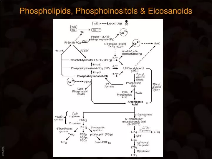
Phospholipids, Phosphoinositols & Eicosanoids Second messenger • released through hydrolysis by phospholipases and/or • generated through the actions of lipid kinases Phospholipases: Phosphatidylinositol-kinases:
Phospholipids, Phosphoinositols & Eicosanoids Phospholipases: • PLA2: • Cytoplasmic form (90 kDa) is regulated through nM Ca++ (Annexins) and phosphorylation; AA specific => signaling function • Secreted form (pancreas, 14 kDa) is also Ca++ dependent (mM range)=> digestive function • PLC: coupled to a variety of (growth factor) receptors: • PLC is activated through GPCRs (Gq) => binding enhances its catalytic activity and in return the GTPase activity of Gq (similar to GAP function in ras signaling) • PLC couples with its SH2 domains directly to growth factor receptors (EGFR, PDGFR) or the TCR, where it is activated through tyrosine phosphorylation
Phospholipids, Phosphoinositols & Eicosanoids Both phospholipases yield finally arachidonic acid (see below), in addition, PLC activity also produces DAG and IP3: • DAG: remains membrane bound; diacylglycerol kinase phosphorylates DAG to generate phosphatidic acid which functions as a substrate for PLA2. Phosphatidyl-serine (PS), Ca++ and DAG activate PKC on the plasma membrane • IP3: see Ca++ signaling!! • Glucocorticoids: inhibit PLA2 by transcriptionally inducing Lipocortin, a protein which binds to PLA2 and blocks its activity. • Phorbol esters: strongest known tumor promotors; mimic DAG => bind PKC and activate it. Also potent activator of Ca2+ influx, MAPK pathway etc.
Phospholipids, Phosphoinositols & Eicosanoids Lipid kinases: • PI3-kinase: • binds to and becomes tyrosine phosphorylated in response to activation of growth factor receptors or immune receptors • 85 kDa regulatory subunit (pY) and a 110 kDa catalytic subunit • regulatory subunit contains SH2 and SH3 domains • PIP3-phosphates can bind to the pleckstrin homology (PH) domain of Akt => Akt activation => phosphorylation of BAD, which dissociates from the antiapoptotic protein bcl-2 => inhibition of apoptosis • Wortmannin: fungal metabolite, potent, irreversible inhibitor of PI3Kinase • Ly290004: synthetic compound, blocks ATP binding site of PI3Kinase
Arachidonic Acid Metabolism • Eicosanoids: collective name for derivatives of arachidonic acid (=5,8,11,14 - eicosatetraenic acid) • AA is mainly generated through the action of PLA2 and DAG-lipase. • Rapidly metabolized by cyclooxygenase and lipoxygenase into prostaglandins and leukotrienes:
Arachidonic Acid Metabolism • Prostaglandins: • First observed in seminal fluid => name • Structure of cyclopentane ring defines letter • Double bonds in side chains account for number • Greek letter refers to the spatial position of the OH-group at C-9 Initial step in PG synthesis catalyzed by PGH-synthase which has dual enzymatic activity: cyclooxygenase (closes ring =>PGG2) and peroxidase (=> 15-OH)
Arachidonic Acid Metabolism Biological functions of PGs: • Vascular toneRelaxation: PGs E1, E2, F2 and I2 Constriction: PGs F2, TxA2 • Platelet aggregationIncrease: PGs E1, TxA2 Decrease: PGs E2, I2 • Uterus toneIncrease: PGs E1, E2, F1 • Bronchial muscleConstriction: PGFs Relaxation: PGEs • Gastric secretionInhibition: PGs E1, E2, I2 • Temperature and painIncrease: PGEs
Arachidonic Acid Metabolism • Leukotrienes: • First found in leucocytes; contain 3 conjugated double bonds • Lipoxygenase generates Hydroperoxyeicosatetraenoic acid (HPETE) • LTC4, D4 and E4 mediate allergic reaction: Slow Reacting Substance of Anaphylaxis (SRS-A) => mediates anaphylactic shock 10,000 fold more potent than histamine!!! => constricts bronchi, dilate blood vessels • LTB4 is a very strong chemoattractant for macrophages
Growth Factor Receptors • Many growth factors (EGF, PDGF, IGF-1, CSF-1, ...) signal through receptors withintrinsic tyrosine kinase activity • Common features: • Large, glycosylated ligand binding domain • Single hydrophobic transmembrane domain • Activation occurs through ligand mediated oligomerization • Undergo ligand induced downregulation by internalization • Cytoplasmic tyrosine kinase domain: • most highly conserved region • GlyXGlyXXGlyX(15-20)Lys Lys is critical for ATP binding - mutation renders receptor kinase inactive, which abrogates all cellular responses => signaling depends on tyrosine phosphorylation of receptor and cytoplasmic substrates • Tyrosine kinase receptors also bind and activate cytoplasmic tyrosine kinases • Autophosphorylation sites: • conserved in the C-term of each receptor class • autophosphorylation does not effect Km of receptor kinase activity • provide docking sites for SH2 domain containg signaling proteins
Growth Factor Receptors • Three subclasses: • Class I: two Cys-rich region in the EC, monomeric ligand EGF-R, erbB2, erbB3, erbB4 (heregulin receptors) • Class II: heterotetrameric: 2 and 2 chains stabilized through S-S bonds: monomeric ligand Insulin-R, IGF-1-R • Class III: Repeats of mmunoglobulin-like structure, dimeric ligand FGF-R, NGF-R, PDGF-R, CSF-1-R, c-kit
Growth Factor Receptors Signaling through Adapter proteins: • grb2: adapter with one SH2 domain which binds PY residue on RTK, and two SH3 domains which bind to • Sos: “Son of Sevenless” (mutation in drosophila prevents development of the R7 photoreceptor cell). Functions as a GEF to facilitate GTP loading of • ras: GTP binding protein, farnesylated; protooncogene, provides a docking site on the plasma membrane for raf. • ras-GAP: negative regulator of groth factor signaling: promotes GTP hydrolysis through the GTPase activity of ras.
Growth Factor Receptors Signaling through Adapter proteins: • raf: ser/thr kinase of the MAPKKK/MEKK family (MEKK does normally NOT phosphorylate MEK, but rather MKK4/7 => Stress pathway); requires context of plasma membrane for activation (mixing GTP-ras and raf in a test tube fails to activate raf) => raf likely to be phosphorylated at the plasma membrane. Activated raf phosphorylates... • MEK: Dual specificity kinase (in case of Stress pathway: SEK) phosphorylates ERKs or MAPKs on tyr and thr -> • ERKs: migrate to the nucleus where they phosphorylate transcription factors such as fos and jun; also feedback loop to other signaling molecules
CytokineReceptors • “Classical” hormones: • produced by cells organized into endocrine organs, • often referred to as “endocrine signal molecules” • target cells usually distant from the site of synthesis • hormone carried by blood stream from producing gland to target cells • signal through receptors coupled to G-proteins (e.g. epinephrine receptor), ion-channels • (e.g. acetylcholine receptor) or receptors with intrinsic enzymatic activity • Cytokines: • single producing and effector cell • only affect target cells in close proximity (autocrine or paracrine) • almost exclusively involved in regulating immunological processes • sometimes subdivided into different groups based on their origin (lymphokines, monokines, interleukines) • often carry several (old) names based on their multiple biological functions: e.g. Lymphocyte Activating Factor (LAF) = Mitogenic Protein (MP) = T Cell Replacing Factor III (TRF-III) = B Cell Activating Factor (BAF) = B cell Differentiation Factor (BDF) = INTERLEUKIN 1
CytokineReceptors • Basic characteristics: • only one copy of encoding gene per haploid cell (~20 different IFN’s, but each encoded by a distinct gene) • genes segmented into 4 or 5 exons (exceptions are IFN and IFN: no introns) • mature protein usually between 8 and 25 kDa • barely any structural resemblances • often N-glycosylated • often form oligomers • some carry signal sequence in precursor • expression is tightly regulated on a transcriptional level • generally pleiotropic • usually highly species specific (up and down) • Multiple (old) classifications: • Based on origin (Lymphokines, Monokines..) • Based on action (inhibitory, stimulating, antiviral, chemotactic...) • Based on composition of receptor (single-chain vs. multi-chain)
CytokineReceptors • Current nomenclature based on structure of receptors: • Type I cytokine receptors = hematopoietin receptor family:receptors contain W-S-X-W-S motif in the C-terminus • IL-2 R, IL-3 R, IL-4 R, IL-5 R, IL-6 R (has also Ig-like domain), IL-7 R, IL-9 R, IL-11 R IL-13 R, IL-15 R, GM-CSF R, EPO R, G-CSF R (has also Ig-like domain) • Type II cytokine receptors = Interferon receptor family:receptors contain IRH1 (200aa extracellular) and IRH2 (50 aa cytoplasmic) domain • IFN R, IFN R • Type III cytokine receptors = TNF receptor family: receptors contain 4 Cys rich regions in extracellular domain • TNF R, TNF=LT R, NGF R (trk), fas, CD40 • Type IV cytokine receptors = Immunoglobulin family: receptors contain an Ig like repeat in the extracellular domain • IL-1 R, M-CSF R (c-fms), SCF R = steel factor R (kit) (tyrosine kinase activity), (IL-6 R) (has also W-S-X-W-S motif), (G-CSF R) • (Chemokine receptors): • C-X-C subgroup (-family): IL-8, PF4, TG • C-C subgroup (-family): RANTES, MCAF, MIP-1
CytokineReceptors Signal Transduction: Receptors lack intrinsic catalytic activity but associate w/ cytosolic enzymes STAT: Signal transducer and activator of transcription • contain SH2 domains • become tyrosine phosphorylated after stimulation • 6 family members • homo or heterodimerize • translocate to nucleus and bind enhancers JAK: Janus kinase • large cytoplasmic tyrosine kinases (130-140 kDa) • NO SH2 or SH3 domains • kinase-like domain
Ser/Thr-kinaseReceptors Transforming Growth Factors = TGFs: • Murine sarcoma virus infected cells can not bind EGF => cells produce a growth factor that competes for EGF binding (=sarcoma growth factor, SGF). • SGF promoted anchorage independent growth (reversible) => first evidence that transformed cells produce their own growth factor! • SGF was found to consist of two subunits: TGF: EGF competitor, binds and signals through the EGF receptor, potent mitogen, but does not support anchorage-independent growth overexpressed in epithelium of psoriasis patients (=> hyperproliferation of keratinocytes) • TGF: acts through distinct receptor, it also is a potent mitogen, but it also does not support anchorage-independent growth (only combination does!) TGF- receptors I and II: • external ligand binding domain and cytosolic serine/threonine kinase activity. • betaglycan: proteoglycan required for TGF binding
Ser/Thr-kinaseReceptors Signal Transduction: SMADs: • 8 members - conserved MH1 and MH2 domain • rapidly phosphorylated in response to TGF (2,3) or BMP (1,5,8) • Smads function in heteromeric complexes • “common” Smad = Smad-4 (Smad-4 required for all Smad signaling) • translocate to the nucleus • phosphorylated by the receptor itself • phosphorylation motif: SSXS • Smad1 potentially also activated by MAPK • Mutations of Smads and /or TGFR are found in 90% of colon cancers
NFkB • originally identified as a transcription factor binding an enhancer (B)in the -light chain immunoglobulin gene • Activated by a variety of (proinflammatory) signals (IL-1, TNF, Phorbol esters...) • Homo-or heterodimer composed of p50 and/or p65 subunits • retained in its inactive form in the cytoplasm by the inhibitory protein IB • dissociation of NFB from IB activates NFB’s DNA binding capabilities • NFB/ IB association is regulated by serine phosphorylation of IB! • Phosphorylated IB does not dissociate from NFB, rather is marked for degradation (NFB activation can be inhibited by protease inhibitors!) • Phosphorylation of IB through IB-kinase (complex >600 kDa) • IKK: two kinase subunits: IKK and IKK homo- or heterodimers IKK (NEMO): no kinase activity, required for complex assembly = regulatory subunit