Download
1 / 1
20 likes | 151 Views
Directing Differentiation of Spinal Progenitor Cells in Hydrogels Sydney A. Geissler , Christine E. Schmidt Dept. of Biomedical Engineering, University of Texas, Austin. Sydney Geissler University of Texas, Austin BME 4.202j sydneygeissler@mail.utexas.edu. Abstract.

E N D
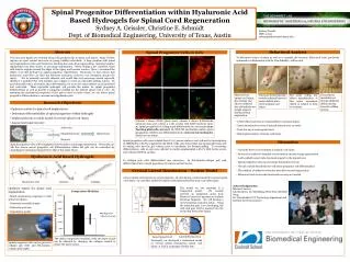
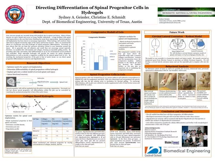
Directing Differentiation of Spinal Progenitor Cells in Hydrogels Sydney A. Geissler, Christine E. Schmidt Dept. of Biomedical Engineering, University of Texas, Austin Sydney Geissler University of Texas, Austin BME 4.202j sydneygeissler@mail.utexas.edu Abstract Compressive Moduli of Gels Future Work Over 200,000 people are currently living with paralysis due to spinal cord injury. Many of these injuries are sport related and occur in young, healthy individuals. A large problem with spinal cord regeneration is the scar that forms, blocking the axons from regenerating. Spinal progenitor cell implantation has been shown to encourage regeneration. When lacking a 3D construct, these cells tend to migrate toward the edges of the injury and become inactive. Here, we examined the effects of a hyaluronic acid (HA) hydrogel on spinal progenitor differentiation. Previously we have shown that HA can limit the astrocyte activation critical to scar formation around the injury. HA is generally non cell adhesive and would thus not encourage axonal regrowth, therefore, we modified HA with laminin and collagen, forming an injectable gelling system. By providing cells with a 3D matrix, their differentiation in an in vivo environment can be predicted and controlled. These injectable hydrogels will provide the matrix for spinal progenitor differentiation as well as provide a supportive scaffold for the injured spinal cord in vivo. By matching the mechanical properties of the gels to that of native tissue, we can direct spinal progenitor differentiation to neurons and oligodendrocytes. Cervical Lateral Contusion – Rodent Model • Optimize modulus for spinal cord implantation. • Match mechanical properties to fetal spinal cord tissue. • Adjust component concentration to optimize modulus. The compressive modulus of the four gel types shows the achievable range with a constant collagen concentration. Laminin and HA incorporation lower the compressive modulus. Adding laminin to the HACol gel does not decrease the modulus further, so HA may play an important role in mediating the compressive strength. • Optimize matrix for spinal cord implantation. • Match mechanical properties to fetal spinal cord tissue. • Determine crosslink density • Determine pore size • Degradation profile HACol Col Col HACol The model we use currently is a transection spinal cord injury model. We recently received an equipment grant from Mission Connect to purchase an Infinite Horizons Impacter. We will develop a cervical lateral contusion injury and use the injectable gels to implant cells into the cavity. Research Objectives Behavioral Analysis • Optimize matrix for spinal cord implantation • Determine differentiability of spinal progenitors within hydrogels • Implant gels into a rodent model of cervical spinal cord injury • Assess functional recovery To determine extent of injury as well as to quantify the recovery, behavioral tests, previously optimized in collaboration with Dr. Tim Schallert, will be used. Spinal Progenitor Cells in Gels LnCol HALnCol LnCol HALnCol Spinal progenitor cells were isolated from E 12.5 mouse embryos and cultured as neurospheres in DMEM/F12 with N2 supplement and bFGF. Cells were dissociated and encapsulated into gels by mixing cells into the gel solution prior to incubation for thermal gelling. To encourage differentiation, cells in gels were cultured in media supplemented with 1% fetal bovine serum and without bFGF for 14 days. Spinal Cord Spinal progenitor cells can be grown in collagen based gels. Col gel (left) and HALnCol gel (right). Lateral Contusion Intact Spinal Cord Virtual Screening Methods for Biomaterial Surface Functionalization + + O4 (green, oligodendrocyte) and DAPI (blue, nuclear) stain for the four different gel types. A portion of implanted cells differentiate into oligodendrocytes(arrows) in all gel types, but fewer oligodendrocytes are seen in LnCol gels, these gels did not promote differentiation toward oligodendrocytes as readily as the other gel types. Scale bars are 50µm. GFAP (red, astrocyte), shows β-III-tubulin (green, neuronal), and DAPI (blue, neuronal) stain in all four gel types. Spinal progenitor cells in Col gels differentiate into astrocytes(arrows). In the three other gel types, a mixed population was present. In HALnCol gels, more neurons (arrowheads) were present than in other gel types. Scale bars are 50µm. Collagen Based Hydrogels Hyaluronic Acid Laminin I Collagen I Astrocytes Oligodendrocytes Neurons "Assessing Forelimb Function after Unilateral Cervical Spinal Cord Injury: Novel Forelimb Tasks Predict Lesion Severity and Recovery“Khaing ZZ, Geissler SA, Jiang S, Milman BD, Aguilar SV, Schmidt C, Schallert T. J. NeurotraumaOnline:2011 Oct 24 We will develop a cervical lateral contusion model of spinal cord injury at C3/C4 in Sprague-Dawley rats. Conclusion and Discussion Spinal progenitor cells will be implanted into the gels to encourage regeneration. Previously our lab has shown neural progenitor cell differentiation within HA gels can be controlled by matching the mechanical properties to that of the native tissue. • The 3D scaffold described here will direct progenitor cell differentiation • Rheological assessment of the gels will reveal their behavior under shear stresses • Oligodendrocyte maturity will be examined with Myelin-Oligodendrocyte Glycoprotein or POP66 (late markers of oligodendrocyte maturation) • Gels will be sectioned to quantify the cellular differentiation and further verify morphological characteristics of the cells in the gels After cervical spinal cord injury, the cylinder test shows unilateral use of forelimbs for weight support during vertical exploration. The postural instability test reveals unilateral deficits during forward walking motion. The pasta eating test allows quantification of fine motor movement deficit as related to time to eat pasta. Vibrissae elicited placing reveals unilateral sensory motor deficits after cervical spinal cord injury Acknowledgements: Mission Connect National Science Foundation Graduate Research Fellowship (grant number: Lab Members: Dr. ZinKhaing (Post Doc) Dr. Tim Schallert (UT Psychology department and Institute for Neuroscience) Adjust mechanical and chemical properties by varying concentrations of collagen, laminin, and HA.