Download
1 / 33
560 likes | 1.36k Views
Screening for phenylketonuria (PKU) – laboratory methods. Péter Monostori. Phe & BH4 metabolism pathway.

E N D
Screening for phenylketonuria (PKU) – laboratory methods Péter Monostori
Phe & BH4 metabolism pathway During the hydroxylation of Phe by Phe hydroxylase (PAH) to form tyrosine (Tyr), tetrahydrobiopterin (BH4) is oxidized to a 4a-hydroxy-BH4 intermediate (when molecular oxygen and iron (Fe2+) are present). q-Dihydrobiopterin
Phe & BH4 metabolism pathway This intermediate is subsequently regenerated back to BH4 via quinonoid (q)-dihydrobiopterin by the enzymes pterin-carbinolamie-4a-dehydratase (PCD) and by the NADH-dependent dihydropteridine reductase (DHPR). Dihydropteridine reductase (DHPR) q-Dihydrobiopterin Pterin-4α-carbinolamine dehydratase (PCD)
Phe & BH4 metabolism pathway GTP cyclohydrolase (GTPCH) BH4 is synthesized from guanosine triphosphate (GTP) by three additional enzymes: GTP cyclohydrolase I (GTPCH), 6-pyruvoyl-tetra-hydropterin synthase (PTPS), and sepiapterin reductase (SR). Mutations in genes coding enzymes for GTPCH, PCD, SR, DHPR and PTPS result in BH4 deficiency. 5% 6-Pyruvoyl-tetrahydrobiopterin synthase (PTPS) 60% Sepiapterin reductase (SR) Dihydropteridine reductase (DHPR) 30% q-Dihydrobiopterin Pterin-4α-carbinolamine dehydratase (PCD) 5%
Which markers can be useful in the diagnosis of PKU and BH4 deficiency? • PKU: • Phe (from DBS) • Tyr (DBS) • Phe/Tyr ratio (DBS) • BH4 deficiency: • Neopterin, biopterin and pterin levels (urine, DBS) • DHPR activity (DBS) • Homovanillic acid (HVA) (liquor) • 5-Hydroxy-indoleacetic acid (5-HIAA) (liquor)
Definitions of positive/negative predictivevalue, sensitivity and specificity • Sensitivity • The proportion of affected subjects that have a positive test result • Specificity • The proportion of unaffected subjects that have a negative test result • Positive predictive value • The chance that a positive test result actually indicates an affected individual • The proportion of „real” positive samples within all positive results • Negative predictive value • The chance that a negative test result actually excludes the disorder • The proportion of „real”negative samples within all negative results
The beginnings… • 1920: A child with developmental delay was born to American parents living in China. No one could help in finding the disease. • The mother wrote a book, describing the symptoms. • The child was later diagnosed as having classical PKU.
Overview of the methods for PKU screening – The Folling-test • 1. In the 1930s: Asbjorn Folling: a mother noticed a strange smell of her mentally retarded child’s urine → Folling analyzed the urine with various tests including the ferric chloride test (for aromatic hydroxyl groups, such as those in ketones): • When ketones are present, urine develops a red-brown colour. • This time the urine turned into dark-green. Folling isolated a substance from the urine which was confirmed to be phenylpyruvate.
The Folling-test • The ferric chloride test (for urine): • not sensitive: usually positive at plasma Phe concentrations above 900 μM (dark green coloration) • not specific: a slightly altered color reaction may be indicative of other metabolic disorders/medication: • maple syrup disease (MSUD) • tyrosinemia • salicylates, L-DOPA metabolites… • traditionally, the reagent was dropped on the diaper of the baby
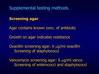
The Guthrie-method • 2. From the 1960s: Robert Guthrie and Ada Susi developed a bacterial inhibition assay, suitable for the screening of PKUfor the first time. • This assay monitors the growth of a mutant strain of Bacillus subtilis with a requirement for exogenous Phe for growth. • DBS samples are placed onto agar plates containing mutant bacteria and an inhibitor. • The sizes of the colonies are assessed after incubation.
The Guthrie-method – principals • The growth of Bacillus subtilis is inhibited by an appropriate amount of β-2-thienylalanine added to the agar. • This inhibition is reversed when a dried blood spot (DBS) containing the blood of a patient with PKU is placed on the agar → Phe in the blood permits the growth of bacteria around the DBS. • The test is positive if the diameter of thegrowth zone is between the 2 mg% (120 μM) and the 4 mg% (240 μM) standard points (marked). • The amount of growth is proportional to the level of Phe in the DBS.
Standards: 2 4 8 16 32 (mg%) The Guthrie-method
The Guthrie-method • Control agar plate without β-2-thienylalanine inhibitor • Rationale: antibiotic therapy can prevent the growth of Bacillus subtilis, resulting in false-negative results • A new blood sample is obtained if a zone with signs of inhibited bacterial growth is found (marked)
The Guthrie-method – characteristics • inexpensive • specific • semiquantitative • not very sensitive: • limit of detection ≈180-240 μM (=3-4 mg%)
Fluorimetric assays • 3. From the 1960s: Fluorimetric assays • McCaman and Robins (1962): • for the determinaton of Phe only • principals: the reaction of Phe, ninhydrin and copper yields a weakly fluorescent product • the fluorescence is increased by the addition of a dipeptide, L-leucyl-L-alanine • Wong, O’Flynn and Inouye (1964): • modified the above method to measure Phe, and added another method to determine Tyr • Ambrose, Ingerson, Garrettson and Cliung(1967): • optimized the Phe-assay by changing several parameters
Fluorimetric assays – characteristics • quantitative • automatization is possible • sensitivity is good: • limit of detection may be as low as 6 μM (0.1 mg%) • not specific (other substances may also yield some degree of fluorescence)
Enzymatic colorimetric assays • 4. From the 1980s: Enzymatic colorimetric assays • Wendel, Hummel and Langenbeck (1989): • for the measurement of Phe using L-phenylalanine dehydrogenase, NAD and a chlorophore • Campbell et al. (1992): • modified the method to reach greater specificity (lower cross-reactivity with Tyr)
Enzymatic colorimetric assays – characteristics • quantitative • automatization is possible • sensitivity is acceptable: • the limit of detection is about 43 μM (0.7 mg%) (higher than that of the fluorescence assay) • specific
Liquid chromatography-tandem mass spectrometry (LC-MS/MS) • 5. From the 1990s: liquid chromatography-tandem mass spectrometry (LC-MS/MS) • allows the simultaneous measurement of a number of disorders of amino acid, organic acid and fatty acid metabolism, including PKU • deuterated internal standards are used • derivatization with butanol-acetyl chloride is employed • selected ratios of the amino acids (or acylcarnitines) are used to help the evaluation
LC-MS/MS assays – characteristics • quantitative • automated • rapid • very sensitive: lower than 1 μM (0.07 mg%) • very specific • the false-positive rate is the lowest, highlighting the advantage of using the Phe/Tyr ratio (Tyr levels are simultaneously measured): • example: parenteral amino acid supplementation or too much blood on the filter paper: Phe ↑, but Phe/Tyr is normal → PKU can be excluded
PKU screening – Blood sampling • primary sample: blood spots dried on filter paper (DBS) • stability of DBS: • ≈10 days at room temperature for amino acids (≈7 days for acylcarnitines) • problems associated with blood sampling: • inappropriate timing • inappropriate technique • delayed delivery • insufficient data on the patient/parent
Problems with blood sampling (DBS) • a) Inappropriate timing of blood sampling (rule: 48-72 h of age; earlier: < 5 days) • the catabolic state associated with birth is the main trigger of most amino acid (incl. Phe) and acylcarnitine elevations in the first few days of life (and not feeding) • (this is not true for galactosaemia and some other disorders) • delayed blood sampling may cause false-negative results
Safe Safe NOT safe Problems with blood sampling (DBS) • b) Inappropriate technique of blood sampling • is mainly responsible for the SD of the MS/MS method • insufficient blood • excess blood • DBS has not dried
Problems with blood sampling (DBS) • c) Delayed delivery of samples • d) Insufficient data on the child/parent • about drugs, parenteral feeding/glucose/middle-chain triglycerides given to the newborn • contact address and telephone number of the parent
The diagnostic value of Phe assays • a positive screening result in a Phe assay is generally sufficient to conclude that some form of hyperphenylalaninemia (PKU, transient hyperphenylalaninemia or BH4 deficiency) is present • confirmation by means of genetic testing or gas chromatography-mass spectrometry (GC/MS) is not essential
Analysis of PKU with (GC/MS): urine samples Plus: 4-hydroxy-phenyllactate, 4-hydroxy-phenylpyruvate, mandelic acid
The diagnostic value of Phe assays (continued) • PKUand BH4 deficiencycannot be distinguished from each other by Phe levels plus Phe/Tyr ratios • for the differential diagnosis of BH4 deficiency: • BH4 loading test, • pterin profile analysis (from urine or DBS), • dihydropteridine reductase (DHPR) activity measurement (from DBS) should be performed
Phe & BH4 metabolism pathway GTP cyclohydrolase (GTPCH) Mutations in genes coding enzymes for BH4 synthesis (GTPCH, PCD, SR), and BH4 recycling (PTPS, DHPR) result in BH4 deficiency. 5% 6-Pyruvoyl-tetrahydrobiopterin synthase (PTPS) 60% Sepiapterin reductase (SR) Dihydropteridine reductase (DHPR) 30% q-Dihydrobiopterin Pterin-4α-carbinolamine dehydratase (PCD) 5%
Diagnosis of BH4 deficiency • 1. BH4 loading test • useful in all forms of BH4 deficiency • single Phe dose plus a single BH4 dose 3 h later • blood sampling: -3; 0; 4; 8; 12; 16; 24 h • (if basal Phe level is low (e.g. < 360 μM), a 24 h Phe loading test may be performed prior to the BH4 test)
Diagnosis of BH4 deficiency • 2. Analysis of pterins with HPLC plus fluorescent or MS/MS detection • Levels of neopterin, biopterin and pterin are measured from urine or DBS. • Chromatographic separation is needed. • Identifies variants: 65-70% of cases. • 3. DHPR activity measurement • Primary sample: DBS • Identifies a single variant: 30-35% of cases.
Pterin levels and DHPR activity in variants of BH4 deficiency
Summary • The ideal method for PKU screening is sensitive, specific, rapid and reliable. • Of the numerous techniques for the measurement of Phe, the Guthrie method, fluorimetric and enzymatic colorimetric assays, and LC-MS/MS (the most recent technique) are used widely for screening purposes. • For the differential diagnosis of hyperphenylalaninemias, BH4 loading test, pterin profile analysis, or measurement of DHPR activity can be performed.