Download
1 / 55
560 likes | 755 Views
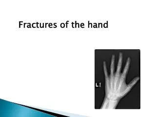
Fractures from Hand to Elbow. Sesamoid bone, found on the palmar surface. Found within the flexor pollicis brevis tendons at the MCP. Constant in position. DP. MP. Biconcave with a median ridge. PP. Concave articular surface. Cancellous bone. MC.

E N D
Sesamoid bone, found on the palmar surface. Found within the flexor pollicis brevis tendons at the MCP. Constant in position. DP MP Biconcave with a median ridge PP Concave articular surface Cancellous bone MC CMC joint Saddle type joint (limited movement) Carpals
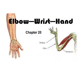
Radiographic Projections DPO DP Hand DP Lateral Finger
Common Fracture sites • Fractures of the Phalanges are more common than the metacarpals • Fractures of the distal phalanx account for over 50% of all phalangeal fractures • Fractures of the middle phalanx are least common, 9-12%
Common Fracture sites Crush, vary from sever to marginal chip fractures Transverse Spiral Avulsion Oblique
Transverse and Longitudinal fractures are less common. • Result of a direct blow DP Marginal Chip fracture 2nd MC 1st
Avulsion of the Flexor Digitorum Profundus Tendon • MOI: • Flexed finger being forcefully extended • E.g. pulling at some ones garment whilst they are pulling away. • Physical signs : inability to Flex finger at the DIP • The tendon retracts proximally to the level of the PIP • With an occasional avulsion fracture at the DIP • A small fracture fragment lying over the volar aspect at the PIP may be seen. • The fragment should not be confused with a fracture of base of the middle phalanx. DIP PIP
History--22-year-old male who comes to the A&E after being injured in a basket ball game. Swelling and deformity to the left index finger Adequacy, Alignment, Bones, Cartilage , Swelling. Joint spaces Which joint ? What type of # ? Where – Dorsal / Volar ? LT. Index
Diagnosis—Intra-articular, avulsion # on dorsal aspect of DIP, at the site of extensor tendon insertion. • MOI--- This injury is due to flexion of a forcibly extended finger, which therefore results in either a tendon injury or a dorsal intra articular avulsion fracture at the dorsal aspect of the distal phalanx. Mallet Finger Or Baseball finger Forced Flexion
Hyperextension at DIP Flexion at PIP Diagnosis--Avulsion fracture on dorsal aspect , At the base of the PIP joint
DIP What is this deformity called ? PIP Boutonniere injury and deformity. (Button hole) Flexed PIP Extended DIP Forced flexion
Struck a wall with fist. Where is the fracture ? Boxers fracture
History--30-year-old female injured while skiing. Swelling and point tenderness over the MC joint. film2 U R film1 Findings--Films 1 and 2 represent a stress view of the left thumb with the normal right thumb also stressed for comparison. There is subluxation of the MCP of the left when compared to the right. There is no evidence of fracture. Diagnosis?
Pole • The ulnar collateral ligament injury is due to a valgus stress. If there is an intraarticular avulsion fracture fragment at the base of the proximal phalanx of the thumb on its ulnar side, the diagnosis is easy. • If you do not see such a fracture fragment, stress views may be required to make the diagnosis. Should be compared with the opposite normal side. Hyperabduction
Described in 1881 by Dr. Edward Bennett; An oblique, intra-articular # at base of the 1st MC (thumb). The fracture extends into the CMC joint with a dorsal subluxation. Due to forced abduction Unstable # pulled by Abductor Pollicis Longus Tendon, in a radial and dorsal direction A small triangular fracture fragment on the volar lip of the base of the MC. This anchored in position by the anterior oblique ligament attached to the volar tubercle of the trapezium.
Rolando`s Fracture: 1910. • A comminuted fracture at the base of the 1st MC, asscociated with dorsal subluxation. • Less common than Bennetts • T or Y type • ORIF
Carpus • Highly complex arrangement of bones and ligaments to allow an infinite variety of movements • During injury stresses are focused on certain sites which lend to the predictability of the site of fracture
Common fracture sites Hamate Scaphoid Triquetral Ulna Styloid
Mechanism of injury • Generally a variation of “foosh” • Injury depends on many variables – • Flexion, extension, rotation, deviation etc. • Results in force focused between radial styloid & capitate across the scaphoid • Proximal row tightly bound to the radius
Frequency of Carpal # Carpal injuries rare in under 12yrs • 70%-80%Scaphoid • 10% dorsal chip # usually Triquetrium • 10% others
Trapezoid 7 Hamate 10 6 Trapezium Capitate 4 5 Scaphoid 9 Triquetral Lunate 3 Pisiform 8 1 Ulna 2 Radius Dorsal Volar
Scaphoid # • 15 – 40 yr of age (rare in children & 60+) • 70% waist • 20% proximal pole • 10% distal pole The scaphoid occupies a vulnerable position, bridging between both rows. With dorsi flexion of hand an wrist, producess greater stresss at the waist of the scaphoid.
Fractures of the distal pole result from compressive forces tansmitted by the index finger and thumb,through the trapezium and trapezoid bones 70 to 80%
Physical Examination Tenderness directly over the scaphoid which lies directly under the anatomical snuff box. There is often swelling in the wrist, and pain with range of motion. Particularly on ulna deviation or making a fist. Tenderness over the ASB is not a specific sign of a scaphoid fracture. 40% with tenderness at this site prove NOT to have a fracture.
Scaphoid views ? Dorsi-palmer Oblique Lateral AP Gripping Zitter 30 30
The scaphoid has a very poor blood supply. It receives its blood supply from the radial artery primarily via lateral volar , dorsal and distal branches. Thus, in one third of waist (mid) fractures, there is diminished blood supply to the proximal fracture fragment This may produce a non-union and lead avascular necrosis . AVN Fractures at each site has specific rates of healing relating to the blood supply of the scaphoid bone
Non-Union Herbert Screw
N UD RD
Fractured Triquetrum 2nd most common site amongst the carpal bones The most common site is a fracture on the dorsal surface of the Triquetral bone. A fractured is generally only seen on the lateral wrist image, always check your laterals for this appearance. This is quite a common fracture. Dorsal radio-triquetral ligament avulsion fracture
Pisiform : acts like a sesamoid bone and lies within the Flexor carpi ulnaris tendon Lateral with 20 degree supination < 1% of carpal bone fractures
Trapezium • Accounts for 3-5 % of the carpal fractures. • Located between the base of the thumb, distal surface of the scaphoid and lateral border of the trapezoid MOI; Abduction of the thumb results in a compression of the radial margin of the trapezium
Fractures - hook of the hamate may be sustained in a fall, more often occurs in sports such as tennis, baseball, and golf, in which a handle sharply impacts the proximal hypothenar palm.Patients who participate in racket sports and present with chronic hand and wrist pain should be suspected of this type of fracture.
Carpal Dislocations • Scapho-Lunate dislocation • Alignment – lines: Gilula`s • Lunate dislocation • Peri-Lunate dislocation • Mid carpal dislocation
Terry Thomas sign ? David Letterman sign Scapho-Lunate dislocation
For normal Alignment on a lateral radiograph: Radius, Lunate and Capitate should all aligned on the lateral projection.
Lunate dislocation The lunate lies outside the carpal boundaries and is thus dislocated
Lunate dislocation Fractures of the Lunate are rare , accounts for < 3% of carpal bone fractures.
Note the overlapping of the proximal and distal carpal rows in addition to the pyramidal appearance of the lunate. Disruption of Gilulas arcs
Peri-lunate Dislocation The lunate remains in it’s normal position but the capitate and neighbouring carpal bones are now out of position. This injury is 2 – 3 times more common than a lunate dislocation and is associated with a Scaphoid fracture 75% of the time.
Mid Carpal dislocation Both the Lunate AND Capitate are dislocate. This is known as a midcarpal dislocation This injury also has a high incidence of associated scaphoid fracture. C L C L R R
MCD LD PLD
Colles`/ Smiths`/ Barton`s A B C C Volar
Abraham Colles` in 1814 • Most common fractures of the forearm. • Age group-adult group over age 40. • More common in females than in males owing to the higher incidence of osteoporosis in women. • 9% of proximal humerus also have colles # • 8% of hip # also have colles #
MOI-- Foosh Mechanism “foosh” compression & tension Compression results in comminution of the dorsal surface • Dorsal displacement of # fragment • +/- ulna styloid # 60% ( ligamentus traction) • 70% intra-articular, 30% extra articular • Disruption of distal radioulnar joint 35%
Smith’s # • Oposite of Colles - “Reverse Colles” • Volar displacement • Fall onto back of hand – wrist supinated • Diagnosis on Lateral X-ray
Smith`s fracture If treated and reduced as a Colles the deformity is maintained
John Barton1838, American surgeon- a fracture ofthe distal end of the radius involving the dorsal rim, with intra-articular extension of the fracture.This injury results from dorsiflexion and pronation of the forearm.Radiographically the fracture is sometimes difficult to distinguish from Colles` fracture, but lateral films show that Barton's fracture does not violate the volar surface of the radius.