Download
1 / 31
320 likes | 383 Views
Elbow, Wrist & Hand Evaluation. BY ABDULLAH RADWAN. Lateral epicondylitis – “tennis elbow” Medial epicondylitis – “golfer’s elbow”, “little league elbow” Hyperextension Sprains DeQuervain’s disease Dislocations Bursitis. Carpal tunnel syndrome Mallet finger Boutonniere deformity

E N D
Elbow, Wrist & Hand Evaluation BY ABDULLAH RADWAN
Lateral epicondylitis – “tennis elbow” Medial epicondylitis – “golfer’s elbow”, “little league elbow” Hyperextension Sprains DeQuervain’s disease Dislocations Bursitis Carpal tunnel syndrome Mallet finger Boutonniere deformity Subungual hematoma Contusions Pathological hand/finger positions (S & R, p.295-300) Fractures Colles’ fx Common Injuries to the Elbow, Wrist, Hand & Fingers
The Elbow – Joints & Movement • Ginglymus or hinge-joint • Humeroulnar joint & Radioulnar joint – 2 interrelated joints • Movements: • Flexion & Extension – primarily between ulna & humerus • 0 to 145 -150
The Elbow – Joints & Movement • Radioulnar joint • Trochoid or pivot-type joint • Syndesmosis – interosseus membrane • Movements: • Supination - 80-90 from neutral • Pronation - 70-90 from neutral
Anatomy ElbowHumerus:TrochleaCapitulum Coronoid Fossa Medial & Lateral EpicondyleRadius:Radial headRadial neckRadial tuberosityRadial FossaUlna:Coronoid ProcessOlecranon ProcessUlna Tuberosity
Elbow LigamentsAnterior ViewAnnularMedial (Ulnar)CollateralLateral (Radial) Collateral
Muscles - Elbow • Flexion • Biceps Brachii • Brachialis • Brachioradialis • **Pronator Teres - weak
Muscles - Elbow • Extension • Triceps Brachii • Anconeus
Muscles - Elbow • Pronation • Pronator Teres • Pronator Quadratus • Brachioradialis
Muscles - Elbow • Supination • Biceps Brachii • Supinator • Brachioradialis
Neuroanatomy • Brachial Plexus – C5, C6, C7, C8, & T1 • Branches: • Radial Nerve (C5, C6, C7 & C8) • Median Nerve (C6 & C7) • Ulnar Nerve (C8 & T1) • Musculocutaneous Nerve (C5 & C6)
DermatomesAnterior View DermatomesPosterior View
Carpal Bones • Concave on palmar side • Bony arch is spanned by transverse carpal & volar ligaments • Creates carpal tunnel • Median nerve & all flexor tendons except flexor carpi ulnaris & palmaris longus pass through carpal tunnel
Anatomical Snuffbox • Extensor pollicis longus (medial side) • Extensor pollicis brevis (lateral side) • Abductor pollicis longus (lateral side) Medial Lateral
Triangular FibrocartilageLigament(TFCC)*extends from ulnar side of distal radius & attaches to ulna @ base of ulnar styloid process*disc provides stability to wrist*major stabilizer of distal radioulnar joint
Joints & Movements • Wrist joint • Condyloid-type • Flexion, extension, abduction (radial deviation), adduction (ulnar deviation) • Motion occurs mostly in proximal carpal row & distal radius • 70°-90° of flexion • 65°-85° of extension • 15°-25° of abduction • 25°-40° of adduction
Joints & Movements • Fingers • Metacarpophalangeal Joint (MCP) • Condyloid • 0°-40° of extension • 85°-100° of flexion • Proximal interphalangeal Joint (PIP) • Ginglymus • Full extension to 90°-120° of flexion • Distal interphalangeal Joint (DIP) • Ginglymus • Flex 80°-90° from full extension
Thumb Joints • 2 joints • Metacarpophalangeal (MCP) • Ginglymus • Full extension into 40°-90° of flexion • Interphalangeal (IP) • Ginglymus • Flex 80°-90° • Carpometacarpal (CMC) joint • Saddle joint • 50°-70° of abduction • Flex 15°-45° & extend 0°-20°
Finger Movement • Middle phalange is reference point to differentiate abduction & adduction • Thumb, index & middle fingers abduct when they move laterally toward radial side of hand • Ring & little fingers abduction when they move medially toward ulnar side of hand • Medial movement of thumb, index & middle fingers toward ulnar side of hand is adduction • Lateral movement of ring & little finger toward radial side of hand is adduction
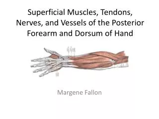
Extrinsic Muscles of Hand Extrinsic muscles of wrist & hand grouped according to function & location • 6 muscles move wrist but not fingers & thumb • 3 wrist flexors • flexor carpi radialis • flexor carpi ulnaris • palmaris longus • 3 wrist extensors • extensor carpi radialis longus • extensor carpi radialis brevis • extensor carpi ulnaris
Extrinsic Muscles of Hand • 9 muscles primary movers of phalanges • Also involved in wrist joint actions • Generally weaker in their wrist actions • Flexors • Flexor digitorum superficialis • Flexor digitorum profundus • Flexor pollicis longus (thumb flexor) • Extensors • Extensor digitorum • Extensor indicis • Extensor digiti minimi • Extensor pollicis longus (thumb extensor) • Extensor pollicis brevis (thumb extensor) • Abductor of thumb & wrist • Abductor pollicis longus
Wrist Abductors & Adductors • Wrist abductors • Generally cross wrist joint anterolaterally & posterolaterally to insert on radial side of hand • Flexor carpi radialis • Extensor carpi radialis longus • Extensor carpi radialis brevis • Abductor pollicis longus • Extensor pollicis longus • Extensor pollicis brevis • Wrist adductors • Cross wrist joint anteromedially & posteromedially to insert on ulnar side of hand • Flexor carpi ulnaris • Extensor carpi ulnaris
Intrinsic Hand Muscles • Intrinsic hand muscles have origins & insertions on bones of hand • Radial side - four muscles of thumb • opponens pollicis • abductor pollicis brevis • flexor pollicis brevis • adductor pollicis • Ulnar side - three muscles of little finger • opponens digiti minimi • abductor digiti minimi • flexor digiti minimi brevis • Remainder of hand - 11 different muscles • 4 lumbricals • 3 palmar interossei • 4 dorsal interossei
Thenar eminence - muscular pad on palmar surface of 1st metacarpal • abductor pollicis brevis • opponens pollicis • flexor pollicis brevis • adductor pollicis • Hypothenar eminence - muscular pad that forms ulnar border on palmar surface • abductor digiti minimi • flexor digiti minimi brevis • opponens digiti minimi
TendonsDorsal AspectExt. Pollicis BrevisExt. Pollicis LongusExt. DigitorumExt. IndicisExt. Digiti Minimi
Elbow & Forearm Evaluation • History • Ask Generic history questions - MOI, ?noises/sensations, Burning/Stinging? • Ask Specific history questions - was your hand planted? Did you fall on an outstretched hand? • Observation • Carrying Angle (> ) • 10-15 5-10 • Cubital Recurvatum • Cubital Valgus • Bony alignment, Soft tissue • Discoloration, swelling, etc.
Elbow & Forearm Evaluation • Palpation • Bony landmarks, Soft tissue • Swelling, crepitus, temperature, etc. • Cubital fossa – brachial artery, median n., musculocutaneous n. • Brachioradialis – lateral border • Pronator teres – medial border • Stress/Special Tests • ROM (AROM, PROM, RROM): - 0-145°; / - normally 0° but can have hyperextension; pronation/supination 170-180° • Valgus/Varus Stress tests • Tinel’s Sign • Compression/Squeeze Test • Neurologic
Wrist & Hand Evaluation • History • Ask Generic history questions - MOI, ?noises/sensations, Burning/Stinging? • Ask Specific history questions - was your hand planted? Did you fall on an outstretched hand? • Observation • Discoloration, swelling • Posture of hand • Deformity, palmar creases, color of skin & fingernails, thenar & hypothenar eminences, thenar webspace • Murphy’s sign, Silverfork deformity, Boutonniere deformity, mallet finger deformity, rotational malalignment, missing knuckle
Wrist & Hand Evaluation • Palpation • Ulna (styloid process), Radius (Lister’s tubercle, styloid process), Carpals, Metacarpals, Phalanges, joints, muscles, ligaments, Carpal Tunnel, Anatomical snuffbox • Temperature, deformity, swelling • Stress/Special Tests • ROM, Grip strength, Pinch test, test ea. joint on a finger • Valgus/Varus Stress tests of all joints, Glide testing of ligaments • Phalen’s Test
Stress/Special Tests continued • Tinel’s Sign • Glide tests – ulnar glide, superior glide, inferior glide • Finkelstein Test • Transverse Compression, Compression Test, Tap or Percussion (Long bone Compression) Test • Watson Test – (Scaphoid shift) • Reflexes • Capillary refill • Pulse
References • Thompson, C. & Floyd, R. T. (2004). Manual of Structural Kinesiology, 15th ed. • Primal 3D Interactive Series • Starkey, C. & Ryan, J. (2003). Orthopedic & Athletic Injury Evaluation Handbook. • Agur, A. & Dalley, A. (2005). Grant’s Atlas of Anatomy. • McDevitt, E. & Roberts, W. (1999). Dorsal Dislocations of the MCP Joint Assessment and Closed Reduction. The Physician & Sportsmedicine, 27(7), p. 75.