Download

1 / 34
730 likes | 1.48k Views
Overview of Stroke. Cerebral Infarction. A Case: Chief Complaint.

E N D
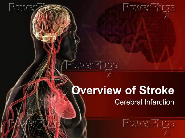
Overview of Stroke Cerebral Infarction
A Case: Chief Complaint • 34 year old female presented to a community hospital with abnormal language. Her husband reported that she had been normal 2 hours earlier at which time the patient is said to have demonstrated shaking of the arms and legs for several seconds of duration. Immediately thereafter the patient was unable to speak and there was paucity of movement on the right side of the body. There was no report of urinary or bowel incontinence and no report of tongue biting.
The Previous Medical History • Migraine Headaches • Frequent Urinary Tract Infections • Multiple episodes of epistaxis • Depression • Miscarriage
Current Medications • Venlafaxine, An antidepressive medication. Works by inhibiting the re-uptake of serotonin, noradrenalin, and dopamine.
Social History • Married • Four living children. G5P5014 • Does not smoke. No history of tobacco use. • No history of recreational or illicit drug use. • No history of alcohol abuse. • No recent travel abroad.
Stroke Epidemiology • First…………..180,000 • Recurrent…….600,000 • Incidence…….780,000/yr =1stk/40s • Prevalence…..6,500,000 • Males…………2,600,000 • Female……….3,900,000
Stroke Mortality • 3rd Leading Cause of Death in USA • 150,000 Deaths Yearly • One of Every 17 Deaths in 2005 • 56,586 Males • 86,993 Females • Death Rate Declined in 2005 i.e. 29.7% to 13.5%
Stroke Morbidity • Leading cause of long term disability • 30% of survivors require assistance with ADL ( activities of daily living) • 20% require assistance to ambulate • 16% must be institutionalized. • Health care and lost income cost approach $41 billion
Stroke by Definition: • An acute on set of neurologic dysfunction caused by impairment of blood flow the region of brain mapping to the impaired function. • Manifest on Brain imaging. • Dysfunction last 24 hours. • If < 24 hours and no signature on brain image: TIA (transient ischemic attack • 0
Classification of Strokes • Hemorrhagic • 15-25 % • ICH • SDH • EDH • Ischemic • 71- 83 % • Embolic • Cardiac Source • Non-cardiac Source • Large Vessel Disease • Small Vessel Disease
Ischemic Stroke Subtypes Large Vessel Small Vessel Embolic (usually Cardioembolic) Thrombotic (usually from Atherosclerotic Cerebrovascular Disease) Microangiopathic Brain Disease Cortical Subcortical
Cardiac Related Stroke Atrial Fibrillation Cardiac Valve disease MI (wall motion abnormality) Septal Aneurysms Patent Foramen Ovale Atrial Septal Defect Dilated Cardiomyopathy
Risk Factors • Hypertension • Heart Disease • Atrial fibrillation • Diabetes • Tobacco • Lipids • Abnormal hematology • OSA • Age • Race/ethnicity • Gender • Family history • genotype
CONGENITAL HYPERCOAGULABLE CONDITIONS • Factor 5 Leiden mutation • G2021A mutation • Antithrombin 3 • Protein C deficiency • Protein S deficiency
Inherited Disorders • Homocystinuria • Fabry’s Disease • Marfan’s Syndrome • Rendu-Osler-Weber Syndrome
34 year old female :Physical Examination • VS: 98.6, 116/71, 23/min, 106/min • Oxygen Saturation 97%, room air • Mute, + commands, neck supple, no bruits, fast RR (-)MRG, clear lungs, abd: benign, extremities: no CCE. • ® Arm>>® Leg Weakness, ® face weakness, (left side normal), Deep Tendon Reflexes absent on the ®, Plantar Response: Up on the ® and Down on the left. Sensation: Normal.
Case laboratory studies • Serum glucose….106 • Sodium…………..142 • Creatinine………...0.7 • WBC………………11,900/cu mm • Platelet count……..258000/cu mm • Hemacrit……….....42% • PT………………….12.9 sec • PTT…………………34.5 sec
Computed Tomography of the Head • Arrival to ER, Airway, Breathing, Circulation, then… • Head CT is crucial in the management of the stroke patient. • The study distinguishes hemorrhagic strokes from ischemic strokes. • The HCT may or may not provide additional diagnostic information • Diffusion weighted MRI: better stroke detection in the first 12 hours.
34 yo female • Dense MCA Sign Head CT Performed 3.5 hours past the onset of stroke symptoms Easter JS et al. N J Med 2010;362:2114-2120.
CT: Thrombosis in the Left Middle Cerebral Artery • Sources of emboli to the brain • Carotid Atherosclerosis • Carotid Dissection • Intracranial Vasculopathy • Atrial Fibrillation • Cardiac Valve Disease • Right-to-Left Cardiac Shunt • Hypercoagulable States
Acute Therapy • Intravenous TPA within 4.5 hrs. of onset • Intra-arterial Thrombolysis within 6 hrs. of onset • Mechanical Embolectomy
Case Patient Acute Therapy • Intravenous Heparin was started. • Patient transferred to tertiary care hospital. • Neurological examination worsened. • Required intubation (protect airway.) • CT angiogram performed.
CT Angiogram of the Brain Filling defect noted in left middle cerebral artery
Case Acute Care Continues • Intra-arterial TPA was administered. • Endovascular mechanical retrieval of clot was performed. • Flow through left MCA was restored. • Right hemiparesis persisted. • Chest x-ray read as right middle lobe pneumonia. • Antibiotic started: patient to ICU.
Chest X-Ray Endotracheal Tube is in place. Right middle lobe infiltrate. Aspiration pneumonia? Or something else?
Additional Studies • Echocardiogram: Suggested Atrial septal defect. • Hypercoagulopathy screen was negative. • Lower extremity venous ultrasound was negative. • Neck CT angiogram negative for dissection.
Putting It All Together • Young female with stroke. • Likely an embolic stroke. • History of nose bleed. • History of miscarriage • Family History of AVM. • Abnormal chest x-ray. • Could be consistent with hereditary hemorrhagic telangiectasia. • Chest CT: Hunt for pulmonary AVM
Contrast CT of the Chest CT Scan of the Torso Obtained 1 Week after Admission. The coronal-plane–formatted CT scan shows an arteriovenous malformation in the lung (arrow).
Cause of this Stroke • Most probably paradoxical through the intrapulmonary shunt created by the pulmonary arteriovenous malformation. • Pulmonary AVM is part and parcel of Rendu-Osler-Weber Syndrome i.e. HHT.
Rendu-Osler-Weber Syndrome • Autosomal Dominant • Telangiectasia of skin mucous membrane, various organs. • Two different Gene Loci identified (a) 9q33-34 and (b) 12q13 • Arises from spontaneous mutations in 30% of cases.
Neurologic Manifestations of HHT • Headache • Dizziness • Seizure • Paradoxical Embolism Stroke • Transient Ischemic Attacks • ICH, SAH • Meningitis • Cerebral abscess
Treatment of HHT • Manage the complications. Notably, our patient walked out of the hospital with improved speech and language. • Early resection of lung AVM or embolization of the fistula. • Periodic Transfusion and Iron Therapy • ASA has been used for platelet sequestration.