Download
1 / 82
820 likes | 895 Views
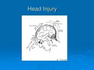
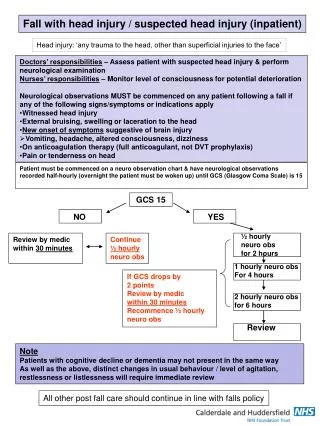
Managing Head Injury at the District Hospital. Pre-test. This patient probably has: Bilateral maxillary fractures Nasal fracture Basal skull fracture Frontal skull fracture. Pre-test.

E N D
Pre-test This patient probably has: • Bilateral maxillary fractures • Nasal fracture • Basal skull fracture • Frontal skull fracture
Pre-test A 70 year old man was a hit by a car. His family bring him to the district hospital. He is unconscious. He does not open his eyes, does not make any sounds, and has no motor response to stimulation. Both pupils are fixed and dilated. What statements are true? • He has a very poor prognosis and will probably die from his head injury. • You should intubate and transfer him immediately to the provincial hospital. • His GCS is 7. • You should check his corneal and gag reflex. • He should receive dilantin to prevent a seizure.
Pre-test A 16 year old boy was in a motor bike accident. He was not wearing a helmet. He is unconscious, airway open and breathing 4 times per minute. His blood pressure is 180/100, pulse 64. His pupils are reactive and not dilated. Which statements are true? • He probably has increased intracranial pressure. • He should be intubated and hyperventilated at about 20 breaths/ minute. • He should receive IV fluid. • He should receive IV antihypertensive medicine. • You should talk to family about sending him to the provincial hospital.
Pre-test Which statements about subdural hematomas are true? Subdural hematomas: • Result from arterial bleeding in the brain • Result from torn veins on surface of brain • Are often associated with diffuse axonal injury • Are most common in children • Have better prognosis than epidural hematomas
Pre-test A young man was admitted to the district hospital yesterday after suffering a head injury. His initial GCS was 11. Today his GCS is 9 and his right pupil is now dilated and not reactive. Which of the following statements are true? • He has developed a delayed intracranial hematoma • He has developed diffuse cerebral edema • You should talk to his family about transfer to the provincial hospital • He should receive mannitol • He should receive D5W IV fluid
Introduction • Severe head injury • Most frequent cause of trauma death • Traffic accident most common cause • Poor prognosis / High mortality and morbidity rates • Difficult decisions to make • Difficult diagnosis with no CT scan • High cost for patient to get transport and have family come and stay at provincial hospital • High cost of CT scan • Patient may have poor outcome • Age is a predictor of outcome • Young patients may do well / Old patients often do poorly
Introduction to Head Injuries • TIME can be CRITICAL • Intracranial Hemorrhage • Progressing Edema • Brain injury from bleed or edema • Increased ICP • Cerebral Hypoxia • Permanent Damage • Severity can be difficult to know
Mechanisms ofHead Injury • Mechanism of Injury • Blunt Injury • Motor vehicle collision / Pedestrian collision • Assault • Fall • Penetrating Injury • Gunshot wound • Stab wound • UXO
Pathophysiology • Blood supply to brain autoregulated as body tries to make sure brain has enough oxygen • 3 types of autoregulation
Pathophysiology • Pressure Autoregulation • If BP , cerebral arteries dilate to keep constant blood supply to brain • Viscousity Autoregulation • If viscousity , cerebral arteries vasodilate • Metabolic Autoregulation • If pO2, cerebral artery vasodilation • If pCO2, cerebral artery vasodilation
Brain Anatomy Intracranial volume • Brain • CSF • Blood vessel volume • Dilatation with high pCO2 • Constriction with low pCO2 Head Trauma - 12
Intracranial Perfusion • Cranial volume fixed • 80% = Cerebrum, cerebellum & brainstem • 12% = Blood vessels & blood • 8% = CSF • Increase in size of one component diminishes size of another • Inability to adjust = increased ICP
Intracranial Perfusion • Compensating Cranial Perfusion for ICP • Compress venous blood vessels • Arterial vasoconstriction in brain • Reduction in free CSF • Push brain into spinal cord • De-compensating Cranial Perfusions for ICP: Increase in ICP • Rise in systemic BP to perfuse brain • Further increase ICP ICP BP
Factors Affecting ICP • Cerebral Edema / Cerebral hematoma • Systemic Systolic BP • Low BP = Poor Cerebral Perfusion • High BP = Increased ICP • Carbon Dioxide & Oxygen levels The Balance: Brain needs sufficient perfusion (O2 delivery), but does not tolerate high ICP
Intracranial Pressure • Role of Systemic Hypotension • Hypotension or under resuscitation will cause higher ICP • Systemic hypotension Brain autoregulation Cerebral vasodilation Increase ICP • If systemic systolic BP < 90 mmHg even 1 occasion, mortality is 1.5 - 2 X higher • AVOID HYPOTENSION IN PATIENTS WITH HEAD INJURY
Intracranial Pressure • Role of Hypoxemia • Brain more sensitive to ischemia after trauma • Hypoxemia Cerebral vasodilation Increase ICP • Higher mortality in patients with hypoxemia and head injury • AVOID HYPOXEMIA IN PATIENTS WITH HEAD INJURY
Intracranial Pressure • Role of Carbon Dioxide • Increase of CO2 • Cerebral Vasodilation • Encourage blood flow • Reduce hypercarbia • Reduce hypoxia • Contributes to ICP • Hyperventilation decreases CO2 in Brain • Cerebral vasoconstriction • Results in cerebral anoxia • AVOID HYPERCARBIA IN PATIENTS WITH HEAD INJURY
Pressure Can Cause Structural Displacement • Increased pressure • Compresses brain tissue • Brain starts to herniate through foramen magnum into brainstem • Compromises blood supply • Signs & Symptoms of pressure on brainstem • Altered mental status • Pupil dilation • Irregular breathing • Bradycardia • Hypertension
Scalp Injuries Scalp wound • Highly vascular, can bleed a lot • Can cause shock in a child • In adult, shock usually indicates another cause • Management • No unstable fracture: Direct pressure & suture • Unstable fracture: Dressings, avoid direct pressure Head Trauma - 21
Skull Fractures Skull fracture • Linear nondisplaced • Depressed • Compound Suspect fracture • Large contusion or darkened swelling Management • Dressing, avoid excess pressure Head Trauma - 22
Skull Fracture • Excess force needed to fracture skull • Types • Linear • Depressed • Open • Impaled Object
Skull Fracture • Depressed skull fracture • Should be referred to provincial hospital for bone elevation if: • Depth of depression more than width of surrounding bone • Open fracture • Neurologic deficit from increased ICP
Basal Skull • Unprotected • Spaces weakenstructure • Relatively easier to fracture
Skull Fracture • Basal Skull Fracture Signs • Battle’s Signs • Retroauricular Ecchymosis • Associated with fracture of auditory canal and lower areas of skull • Raccoon Eyes • Bilateral Periorbital Ecchymosis • Associated with orbital fractures
Basal Skull Fracture Battle’s sign Raccoon eyes Head Trauma - 27
Basal Skull Fracture • Other signs: • Hemotympanum (blood behind tympanic membrane) • Rhinorrhea or Otorrhea (Check fluid for ‘halo’ sign) • NO NG tube because it is possible that tube could pass through fracture and go intracranial
Skull Fracture • Basal Skull Fracture • May tear dura • Permit CSF to drain through nose or ear • Evaluate fluid for “Halo” sign Blood CSF
Forces that cause skull fracture can also cause brain injury. Head Trauma - 30
Classification • Primary • Occur at time of injury • Secondary • Secondary injury caused by factors resulting from the primary injury (ie. cerebral edema)
Brain Injury Primary brain injury • Immediate damage due to force • Coup and contracoup • Occur at time of injury Head Trauma - 32
Primary Brain Injury Types • Coup • Injury at site of impact • Contrecoup • Injury on opposite side from impact
Secondary Brain Injury Response to injury • Swelling of brain • Increased ICP • Decreased blood flow to brain • Perfusion decreases • Cerebral ischemia (hypoxia) Head Trauma - 34
Secondary Brain Injury • Results from hypoxia or decreased perfusion • Response to primary injury • Develops over hours Management • Good prehospital care can help to reduce secondary injury Head Trauma - 35
Signs & Symptoms of Brain Injury • Altered Mental Status • Unconscious • Confusion • Alteration in personality • Amnesia • Retrograde • Antegrade • Cushing’s Reflex • Increased BP • Bradycardia • Erratic respirations • Other • Vomiting • Body temperature changes • Changes in pupil reactivity • Decorticate posturing
Signs & Symptoms of Brain Injury • Origin of Symptoms • Frontal Lobe Injury • Alterations in personality • Occipital Lobe Injury • Visual disturbances • Cortical Disruption • Reduce mental status or Amnesia • Retrograde • Unable to recall events before injury • Antegrade • Unable to recall events after trauma • “Repetitive Questioning” • Focal Deficits • Hemiplegia, Weakness, or Seizures
Primary Brain Injury Categories • Focal • Occur at a specific location in brain • Types • Cerebral Contusion • Intracranial Hemorrhage • Epidural hematoma • Subdural hematoma • Intracerebral Hemorrhage • Diffuse • Concussion • Moderate Diffuse Axonal Injury • Severe Diffuse Axonal Injury
Focal Brain Injury • Cerebral Contusion • Occurs when brain hits bone prominences of skull in blunt trauma • Results from coup-contrecoup injury • Caused by capillary bleeding into brain tissue • 50% of moderate to severe closed head injuries • Symptoms • Prolonged unconsciousness • Severe confusion / amnesia • Possible neurological deficit • Personality changes • Vision changes • Speech changes
Focal Brain InjuryIntracranial Hemorrhage • Epidural Hematoma • 5% of severe head injuries • More common in younger patients • Often from skull fracture that tears middle meningeal artery • Bleeding between dura mater and skull • Rapid bleeding with rapid deterioration
Focal Brain InjuryIntracranial Hemorrhage Epidural hematoma • Level of consciousness • Initial loss of consciousness • “Lucid interval” follows • Consciousness declines within 6 hours • Associated symptoms • Ipsilateral dilated fixed pupil • Signs of increasing ICP • Unconsciousness • Contralateral paralysis • If identified early enough, those who get surgery can do well • Suspect in young patients with possible temporal skull fracture Head Trauma - 41
Focal Brain InjuryIntracranial Hemorrhage • Subdural Hematoma • Bleeding within meninges • Inside dura mater & within subarachnoid space • More often associated with diffuse axonal injury than epidural hematoma • Usually higher force injury • Slow bleeding from veins between brain and venous sinuses • Cerebral atrophy (ie. in elderly or alcoholic patients) puts pts at higher risk
Focal Brain InjuryIntracranial Hemorrhage • Subdural Hematoma • Signs progress over several days • Slow deterioration of mentation • Worse prognosis than epidural hematoma patients • 50 - 90% mortality
Focal Brain InjuryIntracranial Hemorrhage Subdural hematoma • Signs & symptoms • Headache • Fluctuations in level of consciousness • Focal neurological signs (ie. weakness of one side or limb, slurred speech) Head Trauma - 44
Focal Brain Injury Intracerebral hemorrhage • Arterial or venous • Surgery is often not helpful • Level of consciousness • Alterations common • Associated symptoms • Varies with region and degree • Pattern similar to stroke • Headache and vomiting Head Trauma - 45
Diffuse Brain Injury • Due to stretching forces placed on individual nerve cells • Injury distributed throughout brain • Types • Concussion • Moderate Diffuse Axonal Injury • Severe Diffuse Axonal Injury
Diffuse Brain Injury • Diffuse axonal injury • Generalized edema • No structural lesion • Most common injury fromsevere blunt head trauma Head Trauma - 47
Diffuse Brain InjuryConcussion • Mild form of Diffuse Axonal Injury (DAI) • Nerve dysfunction without anatomic damage • CT scan usually normal • Short episode of: • Loss of consciousness • Confusion • Event amnesia • Headache, dizziness, nausea • GCS usually 14 or 15 • Usually good recovery
Diffuse Brain InjuryModerate Diffuse Axonal Injury • Same mechanism as concussion, but more severe bruising of brain tissue • Signs & Symptoms • Prolonged unconsciousness • Persistent confusion • Loss of concentration • Retrograde & antegrade amnesia • Mood or personality changes
Diffuse Brain InjurySevere Diffuse Axonal Injury • Significant mechanical disruption of nerve cells • Cerebral hemispheres AND brainstem • High mortality rate • Signs & Symptoms • Prolonged unconsciousness • Cushing’s reflex • Decorticate or Decerebrate posturing • Dilated fixed pupils